3
views
0
recommends
+1 Recommend
0 collections
    0
    shares
      • Record: found
      • Abstract: found
      • Article: found
      Is Open Access

      Effect of Adding Nano Size Silica on Setting Time and Porosity of Mineral Trioxide Aggregate

      research-article

      Read this article at

      Bookmark
          There is no author summary for this article yet. Authors can add summaries to their articles on ScienceOpen to make them more accessible to a non-specialist audience.

          Abstract

          Introduction:

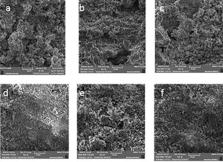
          The aim of this study was to evaluate the effect of addition of nano-silica (SiO 2) to mineral trioxide aggregate (MTA) on its setting time and porosity.

          Methods and Materials:

          The concentration 8% of nano-silica were prepared and added to the MTA powder. After mixing with water the setting time and porosity were evaluated and compared with pure MTA. Statistical analysis was performed using the t-test. The level of significance was set at 0.001.

          Results:

          The mean setting time of MTA+8% nano-silica (9.8±0.78) was significantly lower than MTA (23.3±2.16) ( P<0.001). Also the mean porosity by imbibition method in MTA+8% nano-silica (23.49±0.48) was significantly higher than MTA (15.69±2.10) ( P<0.001). There was no significant difference in mean porosity by scanning electron microscope (SEM) method in MTA+8% nano-silica (31.26±10.73) and MTA (32.74±5.26) ( P>0.001).

          Conclusion:

          This in vitro study showed us an addition of 8% of nano-silica to MTA reduced the setting time. Although evaluation by imbibition test showed increasing of porosity in nano-silica MTA compared with pure MTA.

          Related collections

          Most cited references34

          • Record: found
          • Abstract: found
          • Article: not found

          Mineral trioxide aggregate: a comprehensive literature review--Part III: Clinical applications, drawbacks, and mechanism of action.

          Mineral trioxide aggregate (MTA) has been recommended for various uses in endodontics. Two previous publications provided a comprehensive list of articles from November 1993-September 2009 regarding the chemical and physical properties, sealing ability, antibacterial activity, leakage, and biocompatibility of MTA. The purpose of Part III of this literature review is to present a comprehensive list of articles regarding animal studies, clinical applications, drawbacks, and mechanism of action of MTA. A review of the literature was performed by using electronic and hand-searching methods for the clinical applications of MTA in experimental animals and humans as well as its drawbacks and mechanism of action from November 1993-September 2009. MTA is a promising material for root-end filling, perforation repair, vital pulp therapy, and apical barrier formation for teeth with necrotic pulps and open apexes. Despite the presence of numerous case reports and case series regarding these applications, there are few designed research studies regarding clinical applications of this material. MTA has some known drawbacks such as a long setting time, high cost, and potential of discoloration. Hydroxyapatite crystals form over MTA when it comes in contact with tissue synthetic fluid. This can act as a nidus for the formation of calcified structures after the use of this material in endodontic treatments. On the basis of available information, it appears that MTA is the material of choice for some clinical applications. More clinical studies are needed to confirm its efficacy compared with other materials. Copyright (c) 2010. Published by Elsevier Inc.
            Bookmark
            • Record: found
            • Abstract: found
            • Article: not found

            Mineral trioxide aggregate: a comprehensive literature review--Part I: chemical, physical, and antibacterial properties.

            An ideal orthograde or retrograde filling material should seal the pathways of communication between the root canal system and its surrounding tissues. It should also be nontoxic, noncarcinogenic, nongenotoxic, biocompatible, insoluble in tissue fluids, and dimensionally stable. Mineral trioxide aggregate (MTA) was developed and recommended initially because existing root-end filling materials did not have these "ideal" characteristics. MTA has also been recommended for pulp capping, pulpotomy, apical barrier formation in teeth with open apexes, repair of root perforations, and root canal filling. Since MTA's introduction in 1993, numerous studies have been published regarding various aspects of this material. The aim of Part I of this literature review is to present investigations regarding the chemical, physical, and antibacterial properties of MTA. A review of the literature was performed by using electronic and hand-searching methods for the chemical and physical properties and antibacterial activity of MTA from November 1993-September 2009. There are many published reports regarding the chemical, physical, and antibacterial properties of MTA. Our search showed that MTA is composed of calcium, silica, and bismuth. It has a long setting time, high pH, and low compressive strength. It possesses some antibacterial and antifungal properties, depending on its powder-to-liquid ratio. MTA is a bioactive material that influences its surrounding environment.
              Bookmark
              • Record: found
              • Abstract: found
              • Article: not found

              Biomineralization ability and interaction of mineral trioxide aggregate and white portland cement with dentin in a phosphate-containing fluid.

              Mineral trioxide aggregate (MTA) has been shown to be bioactive because of its ability to produce biologically compatible carbonated apatite. This study analyzed the interaction of MTA and white Portland cement with dentin after immersion in phosphate-buffered saline (PBS). Dentin disks with standardized cavities were filled with ProRoot MTA, MTA Branco, MTA BIO, white Portland cement + 20% bismuth oxide (PC1), or PC1 + 10% of calcium chloride (PC2) and immersed in 15 mL of PBS for 2 months. The precipitates were weighed and analyzed by scanning electron microscopy (SEM) and x-ray diffraction. The calcium ion release and pH of the solutions were monitored at 5, 15, 25, and 35 days. The samples were processed for SEM observations. Data were analyzed by using analysis of variance or Kruskall-Wallis tests. Our findings revealed the presence of amorphous calcium phosphate precipitates with different morphologies. The apatite formed by the cement-PBS system was deposited within collagen fibrils, promoting controlled mineral nucleation on dentin, observed as the formation of an interfacial layer with tag-like structures. All the cements tested were bioactive. The cements release some of their components in PBS, triggering the initial precipitation of amorphous calcium phosphates, which act as precursors during the formation of carbonated apatite. This spontaneous precipitation promotes a biomineralization process that leads to the formation of an interfacial layer with tag-like structures at the cement-dentin interface.
                Bookmark

                Author and article information

                Journal
                Iran Endod J
                Iran Endod J
                IEJ
                Iranian Endodontic Journal
                Iranian Center for Endodontic Research (Tehran, Iran )
                1735-7497
                2008-2746
                Summer 2019
                : 14
                : 3
                : 197-201
                Affiliations
                [a ] Dental Research Center, Mashhad University of Medical Sciences, Mashhad, Iran;
                [b ] Private Practice, Mashhad, Iran;
                [c ] Department of Chemical Engineering, Faculty of Engineering, Ferdowsi University of Mashhad, Mashhad, Iran;
                [d ] Department of Endodontics, Mashhad University of Medical Sciences, Mashhad, Iran
                Author notes
                [* ]Corresponding author: Ehsan Ashrafpour, Postgraduate Student of Endodontics, Department of Endodontics Faculty of Dentistry, Mashhad University of Medical Sciences, Mashhad, Iran. Tel: +98-915 3158324 Email: ehsan.ashrafpour@gmail.com
                Article
                10.22037/iej.v14i3.23010
                9940024
                36814945
                262e1a78-335d-4860-a259-4b1b46f12bdd

                This work is licensed under the terms of the Creative Commons Attribution-NonCommercial-ShareAlike 4.0 International.( https://creativecommons.org/licenses/by-nc-sa/4.0/)

                History
                : 28 December 2018
                : 3 June 2019
                : 17 June 2019
                Categories
                Original Article

                Dentistry
                mineral trioxide aggregate,nano-silica; porosity,setting time
                Dentistry
                mineral trioxide aggregate, nano-silica; porosity, setting time

                Comments

                Comment on this article