9
views
0
recommends
+1 Recommend
0 collections
    0
    shares
      • Record: found
      • Abstract: found
      • Article: found
      Is Open Access

      Antidromic Spike Propagation and Dissimilar Expression of P2X, 5-HT, and TRPV1 Channels in Peripheral vs. Central Sensory Axons in Meninges

      brief-report

      Read this article at

      Bookmark
          There is no author summary for this article yet. Authors can add summaries to their articles on ScienceOpen to make them more accessible to a non-specialist audience.

          Abstract

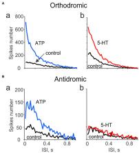
          Background: The terminal branches of the trigeminal nerve in meninges are supposed to be the origin site of migraine pain. The main function of these peripheral sensory axons is the initiation and propagation of spikes in the orthodromic direction to the second order neurons in the brainstem. The stimulation of the trigeminal ganglion induces the release of the neuropeptide CGRP in meninges suggesting the antidromic propagation of excitation in these fibers. However, the direct evidence on antidromic spike traveling in meningeal afferents is missing.

          Methods: By recording of spikes from peripheral or central parts of the trigeminal nerve in rat meninges, we explored their functional activity and tested the expression of ATP-, serotonin-, and capsaicin-gated receptors in the distal vs. proximal parts of these nerves.

          Results: We show the significant antidromic propagation of spontaneous spikes in meningeal nerves which was, however, less intense than the orthodromic nociceptive traffic due to higher number of active fibers in the latter. Application of ATP, serotonin and capsaicin induced a high frequency nociceptive firing in peripheral processes while, in central parts, only ATP and capsaicin were effective. Disconnection of nerve from trigeminal ganglion dramatically reduced the tonic antidromic activity and attenuated the excitatory action of ATP.

          Conclusion: Our data indicate the bidirectional nociceptive traffic and dissimilar expression of P2X, 5-HT and TRPV1 receptors in proximal vs. distal parts of meningeal afferents, which is important for understanding the peripheral mechanisms of migraine pain.

          Related collections

          Most cited references45

          • Record: found
          • Abstract: found
          • Article: not found

          Endothelial cation channel PIEZO1 controls blood pressure by mediating flow-induced ATP release.

          Arterial blood pressure is controlled by vasodilatory factors such as nitric oxide (NO) that are released from the endothelium under the influence of fluid shear stress exerted by flowing blood. Flow-induced endothelial release of ATP and subsequent activation of Gq/G11-coupled purinergic P2Y2 receptors have been shown to mediate fluid shear stress-induced stimulation of NO formation. However, the mechanism by which fluid shear stress initiates these processes is unclear. Here, we have shown that the endothelial mechanosensitive cation channel PIEZO1 is required for flow-induced ATP release and subsequent P2Y2/Gq/G11-mediated activation of downstream signaling that results in phosphorylation and activation of AKT and endothelial NOS. We also demonstrated that PIEZO1-dependent ATP release is mediated in part by pannexin channels. The PIEZO1 activator Yoda1 mimicked the effect of fluid shear stress on endothelial cells and induced vasorelaxation in a PIEZO1-dependent manner. Furthermore, mice with induced endothelium-specific PIEZO1 deficiency lost the ability to induce NO formation and vasodilation in response to flow and consequently developed hypertension. Together, our data demonstrate that PIEZO1 is required for the regulation of NO formation, vascular tone, and blood pressure.
            Bookmark
            • Record: found
            • Abstract: found
            • Article: not found

            Origin of pain in migraine: evidence for peripheral sensitisation.

            Migraine is the most common neurological disorder, and much has been learned about its mechanisms in recent years. However, the origin of painful impulses in the trigeminal nerve is still uncertain. Despite the attention paid recently to the role of central sensitisation in migraine pathophysiology, in our view, neuronal hyperexcitability depends on activation of peripheral nociceptors. Although the onset of a migraine attack might take place in deep-brain structures, some evidence indicates that the headache phase depends on nociceptive input from perivascular sensory nerve terminals. The input from arteries is probably more important than the input from veins. Several studies provide evidence for input from extracranial, dural, and pial arteries but, likewise, there is also evidence against all three of these locations. On balance, afferents are most probably excited in all three territories or the importance of individual territories varies from patient to patient. We suggest that migraine can be explained to patients as a disorder of the brain, and that the headache originates in the sensory fibres that convey pain signals from intracranial and extracranial blood vessels.
              Bookmark
              • Record: found
              • Abstract: found
              • Article: not found

              Molecular mechanisms of nociception.

              The sensation of pain alerts us to real or impending injury and triggers appropriate protective responses. Unfortunately, pain often outlives its usefulness as a warning system and instead becomes chronic and debilitating. This transition to a chronic phase involves changes within the spinal cord and brain, but there is also remarkable modulation where pain messages are initiated - at the level of the primary sensory neuron. Efforts to determine how these neurons detect pain-producing stimuli of a thermal, mechanical or chemical nature have revealed new signalling mechanisms and brought us closer to understanding the molecular events that facilitate transitions from acute to persistent pain.
                Bookmark

                Author and article information

                Contributors
                Journal
                Front Cell Neurosci
                Front Cell Neurosci
                Front. Cell. Neurosci.
                Frontiers in Cellular Neuroscience
                Frontiers Media S.A.
                1662-5102
                15 January 2021
                2020
                : 14
                : 623134
                Affiliations
                [1] 1Department of Human and Animal Physiology, Institute of Fundamental Medicine and Biology, Kazan Federal University , Kazan, Russia
                [2] 2A.I. Virtanen Institute for Molecular Sciences, Faculty of Health Sciences, University of Eastern Finland , Kuopio, Finland
                Author notes

                Edited by: David J. Adams, University of Wollongong, Australia

                Reviewed by: Gábor Jancsó, University of Szeged, Hungary; Karl Messlinger, University of Erlangen Nuremberg, Germany

                *Correspondence: Rashid Giniatullin rashid.giniatullin@ 123456uef.fi

                This article was submitted to Cellular Neurophysiology, a section of the journal Frontiers in Cellular Neuroscience

                †These authors share first authorship

                Article
                10.3389/fncel.2020.623134
                7845021
                33519387
                2912db64-de0a-4043-bac7-2759eb3f8f32
                Copyright © 2021 Gafurov, Koroleva and Giniatullin.

                This is an open-access article distributed under the terms of the Creative Commons Attribution License (CC BY). The use, distribution or reproduction in other forums is permitted, provided the original author(s) and the copyright owner(s) are credited and that the original publication in this journal is cited, in accordance with accepted academic practice. No use, distribution or reproduction is permitted which does not comply with these terms.

                History
                : 29 October 2020
                : 17 December 2020
                Page count
                Figures: 4, Tables: 0, Equations: 0, References: 45, Pages: 9, Words: 6227
                Funding
                Funded by: Russian Foundation for Basic Research 10.13039/501100002261
                Funded by: Kazan Federal University 10.13039/501100012528
                Categories
                Cellular Neuroscience
                Brief Research Report

                Neurosciences
                migraine,meninges,trigeminal nerve,excitability,atp,5-ht,trpv1 receptor
                Neurosciences
                migraine, meninges, trigeminal nerve, excitability, atp, 5-ht, trpv1 receptor

                Comments

                Comment on this article