25
views
0
recommends
+1 Recommend
0 collections
    0
    shares
      • Record: found
      • Abstract: found
      • Article: found
      Is Open Access

      Intermediate Filaments at the Junction of Mechanotransduction, Migration, and Development

      review-article

      Read this article at

      Bookmark
          There is no author summary for this article yet. Authors can add summaries to their articles on ScienceOpen to make them more accessible to a non-specialist audience.

          Abstract

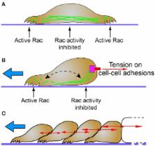
          Mechanically induced signal transduction has an essential role in development. Cells actively transduce and respond to mechanical signals and their internal architecture must manage the associated forces while also being dynamically responsive. With unique assembly-disassembly dynamics and physical properties, cytoplasmic intermediate filaments play an important role in regulating cell shape and mechanical integrity. While this function has been recognized and appreciated for more than 30 years, continually emerging data also demonstrate important roles of intermediate filaments in cell signal transduction. In this review, with a particular focus on keratins and vimentin, the relationship between the physical state of intermediate filaments and their role in mechanotransduction signaling is illustrated through a survey of current literature. Association with adhesion receptors such as cadherins and integrins provides a critical interface through which intermediate filaments are exposed to forces from a cell's environment. As a consequence, these cytoskeletal networks are posttranslationally modified, remodeled and reorganized with direct impacts on local signal transduction events and cell migratory behaviors important to development. We propose that intermediate filaments provide an opportune platform for cells to both cope with mechanical forces and modulate signal transduction.

          Related collections

          Most cited references200

          • Record: found
          • Abstract: found
          • Article: not found

          Focal Contacts as Mechanosensors

          The transition of cell–matrix adhesions from the initial punctate focal complexes into the mature elongated form, known as focal contacts, requires GTPase Rho activity. In particular, activation of myosin II–driven contractility by a Rho target known as Rho-associated kinase (ROCK) was shown to be essential for focal contact formation. To dissect the mechanism of Rho-dependent induction of focal contacts and to elucidate the role of cell contractility, we applied mechanical force to vinculin-containing dot-like adhesions at the cell edge using a micropipette. Local centripetal pulling led to local assembly and elongation of these structures and to their development into streak-like focal contacts, as revealed by the dynamics of green fluorescent protein–tagged vinculin or paxillin and interference reflection microscopy. Inhibition of Rho activity by C3 transferase suppressed this force-induced focal contact formation. However, constitutively active mutants of another Rho target, the formin homology protein mDia1 (Watanabe, N., T. Kato, A. Fujita, T. Ishizaki, and S. Narumiya. 1999. Nat. Cell Biol. 1:136–143), were sufficient to restore force-induced focal contact formation in C3 transferase-treated cells. Force-induced formation of the focal contacts still occurred in cells subjected to myosin II and ROCK inhibition. Thus, as long as mDia1 is active, external tension force bypasses the requirement for ROCK-mediated myosin II contractility in the induction of focal contacts. Our experiments show that integrin-containing focal complexes behave as individual mechanosensors exhibiting directional assembly in response to local force.
            Bookmark
            • Record: found
            • Abstract: found
            • Article: not found

            Flexural rigidity of microtubules and actin filaments measured from thermal fluctuations in shape

            Microtubules are long, proteinaceous filaments that perform structural functions in eukaryotic cells by defining cellular shape and serving as tracks for intracellular motor proteins. We report the first accurate measurements of the flexural rigidity of microtubules. By analyzing the thermally driven fluctuations in their shape, we estimated the mean flexural rigidity of taxol-stabilized microtubules to be 2.2 x 10(-23) Nm2 (with 6.4% uncertainty) for seven unlabeled microtubules and 2.1 x 10(-23) Nm2 (with 4.7% uncertainty) for eight rhodamine-labeled microtubules. These values are similar to earlier, less precise estimates of microtubule bending stiffness obtained by modeling flagellar motion. A similar analysis on seven rhodamine-phalloidin- labeled actin filaments gave a flexural rigidity of 7.3 x 10(-26) Nm2 (with 6% uncertainty), consistent with previously reported results. The flexural rigidity of these microtubules corresponds to a persistence length of 5,200 microns showing that a microtubule is rigid over cellular dimensions. By contrast, the persistence length of an actin filament is only approximately 17.7 microns, perhaps explaining why actin filaments within cells are usually cross-linked into bundles. The greater flexural rigidity of a microtubule compared to an actin filament mainly derives from the former's larger cross-section. If tubulin were homogeneous and isotropic, then the microtubule's Young's modulus would be approximately 1.2 GPa, similar to Plexiglas and rigid plastics. Microtubules are expected to be almost inextensible: the compliance of cells is due primarily to filament bending or sliding between filaments rather than the stretching of the filaments themselves.
              Bookmark
              • Record: found
              • Abstract: found
              • Article: not found

              Collective epithelial migration and cell rearrangements drive mammary branching morphogenesis.

              Epithelial organs are built through the movement of groups of interconnected cells. We observed cells in elongating mammary ducts reorganize into a multilayered epithelium, migrate collectively, and rearrange dynamically, all without forming leading cellular extensions. Duct initiation required proliferation, Rac, and myosin light-chain kinase, whereas repolarization to a bilayer depended on Rho kinase. We observed that branching morphogenesis results from the active motility of both luminal and myoepithelial cells. Luminal epithelial cells advanced collectively, whereas myoepithelial cells appeared to restrain elongating ducts. Significantly, we observed that normal epithelium and neoplastic hyperplasias are organized similarly, suggesting common mechanisms of epithelial growth.
                Bookmark

                Author and article information

                Contributors
                Journal
                Front Cell Dev Biol
                Front Cell Dev Biol
                Front. Cell Dev. Biol.
                Frontiers in Cell and Developmental Biology
                Frontiers Media S.A.
                2296-634X
                14 September 2017
                2017
                : 5
                : 81
                Affiliations
                Department of Biological Sciences, Rutgers University-Newark Newark, NJ, United States
                Author notes

                Edited by: Takaaki Matsui, Nara Institute of Science and Technology, Japan

                Reviewed by: Kensaku Mizuno, Tohoku University, Japan; Thomas Magin, Leipzig University, Germany

                *Correspondence: Gregory F. Weber gregory.weber@ 123456rutgers.edu

                This article was submitted to Cell Adhesion and Migration, a section of the journal Frontiers in Cell and Developmental Biology

                Article
                10.3389/fcell.2017.00081
                5603733
                28959689
                2bf9508c-2dc6-4d70-b3e5-ffbef483853d
                Copyright © 2017 Sanghvi-Shah and Weber.

                This is an open-access article distributed under the terms of the Creative Commons Attribution License (CC BY). The use, distribution or reproduction in other forums is permitted, provided the original author(s) or licensor are credited and that the original publication in this journal is cited, in accordance with accepted academic practice. No use, distribution or reproduction is permitted which does not comply with these terms.

                History
                : 30 June 2017
                : 30 August 2017
                Page count
                Figures: 2, Tables: 3, Equations: 0, References: 232, Pages: 19, Words: 17577
                Funding
                Funded by: Eunice Kennedy Shriver National Institute of Child Health and Human Development 10.13039/100009633
                Award ID: R15HD084254
                Categories
                Cell and Developmental Biology
                Review

                intermediate filaments,development,migration,mechanotransduction,tension,keratin,vimentin

                Comments

                Comment on this article