3
views
0
recommends
+1 Recommend
0 collections
    0
    shares
      • Record: found
      • Abstract: found
      • Article: found
      Is Open Access

      A Profound Basic Characterization of eIFs in Gliomas: Identifying eIF3I and 4H as Potential Novel Target Candidates in Glioma Therapy

      research-article

      Read this article at

      Bookmark
          There is no author summary for this article yet. Authors can add summaries to their articles on ScienceOpen to make them more accessible to a non-specialist audience.

          Abstract

          Simple Summary

          Gliomas are brain tumors with currently limited therapy options. Glioma growth and proliferation is regulated by the mTOR pathway together with eukaryotic initiation factors (eIFs). In this work we show a profound basic characterization of eIFs in human gliomas and demonstrate increased mRNA and protein expressions of several eIFs in gliomas compared to healthy control brain tissue. Moreover, increased eIF3I and eIF4H levels seem to have a negative influence on the survival of patients. Our work suggests eIF3I and eIF4H as potential targets for future glioma therapy.

          Abstract

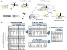
          Glioblastoma (GBM) is an utterly devastating cerebral neoplasm and current therapies only marginally improve patients’ overall survival (OS). The PI3K/AKT/mTOR pathway participates in gliomagenesis through regulation of cell growth and proliferation. Since it is an upstream regulator of the rate-limiting translation initiation step of protein synthesis, controlled by eukaryotic initiation factors (eIFs), we aimed for a profound basic characterization of 17 eIFs to identify potential novel therapeutic targets for gliomas. Therefore, we retrospectively analyzed expressions of mTOR-related proteins and eIFs in human astrocytoma samples (WHO grades I–IV) and compared them to non-neoplastic cortical control brain tissue (CCBT) using immunoblot analyses and immunohistochemistry. We examined mRNA expression using qRT-PCR and additionally performed in silico analyses to observe the influence of eIFs on patients’ survival. Protein and mRNA expressions of eIF3B, eIF3I, eIF4A1, eIF4H, eIF5 and eIF6 were significantly increased in high grade gliomas compared to CCBT and partially in low grade gliomas. However, short OS was only associated with high eIF3I gene expression in low grade gliomas, but not in GBM. In GBM, high eIF4H gene expression significantly correlated with shorter patient survival. In conclusion, we identified eIF3I and eIF4H as the most promising targets for future therapy for glioma patients.

          Related collections

          Most cited references40

          • Record: found
          • Abstract: found
          • Article: not found

          Analysis of relative gene expression data using real-time quantitative PCR and the 2(-Delta Delta C(T)) Method.

          The two most commonly used methods to analyze data from real-time, quantitative PCR experiments are absolute quantification and relative quantification. Absolute quantification determines the input copy number, usually by relating the PCR signal to a standard curve. Relative quantification relates the PCR signal of the target transcript in a treatment group to that of another sample such as an untreated control. The 2(-Delta Delta C(T)) method is a convenient way to analyze the relative changes in gene expression from real-time quantitative PCR experiments. The purpose of this report is to present the derivation, assumptions, and applications of the 2(-Delta Delta C(T)) method. In addition, we present the derivation and applications of two variations of the 2(-Delta Delta C(T)) method that may be useful in the analysis of real-time, quantitative PCR data. Copyright 2001 Elsevier Science (USA).
            Bookmark
            • Record: found
            • Abstract: found
            • Article: not found

            NIH Image to ImageJ: 25 years of image analysis

            For the past twenty five years the NIH family of imaging software, NIH Image and ImageJ have been pioneers as open tools for scientific image analysis. We discuss the origins, challenges and solutions of these two programs, and how their history can serve to advise and inform other software projects.
              Bookmark
              • Record: found
              • Abstract: found
              • Article: found

              The 2016 World Health Organization Classification of Tumors of the Central Nervous System: a summary.

              The 2016 World Health Organization Classification of Tumors of the Central Nervous System is both a conceptual and practical advance over its 2007 predecessor. For the first time, the WHO classification of CNS tumors uses molecular parameters in addition to histology to define many tumor entities, thus formulating a concept for how CNS tumor diagnoses should be structured in the molecular era. As such, the 2016 CNS WHO presents major restructuring of the diffuse gliomas, medulloblastomas and other embryonal tumors, and incorporates new entities that are defined by both histology and molecular features, including glioblastoma, IDH-wildtype and glioblastoma, IDH-mutant; diffuse midline glioma, H3 K27M-mutant; RELA fusion-positive ependymoma; medulloblastoma, WNT-activated and medulloblastoma, SHH-activated; and embryonal tumour with multilayered rosettes, C19MC-altered. The 2016 edition has added newly recognized neoplasms, and has deleted some entities, variants and patterns that no longer have diagnostic and/or biological relevance. Other notable changes include the addition of brain invasion as a criterion for atypical meningioma and the introduction of a soft tissue-type grading system for the now combined entity of solitary fibrous tumor / hemangiopericytoma-a departure from the manner by which other CNS tumors are graded. Overall, it is hoped that the 2016 CNS WHO will facilitate clinical, experimental and epidemiological studies that will lead to improvements in the lives of patients with brain tumors.
                Bookmark

                Author and article information

                Contributors
                Role: Academic Editor
                Journal
                Cancers (Basel)
                Cancers (Basel)
                cancers
                Cancers
                MDPI
                2072-6694
                23 March 2021
                March 2021
                : 13
                : 6
                : 1482
                Affiliations
                [1 ]Diagnostic & Research Center for Molecular BioMedicine, Department of Neuropathology, Diagnostic and Research Institute of Pathology, Medical University Graz, Neue Stiftingtalstrasse 6, 8010 Graz, Austria; stefanie.krassnig@ 123456medunigraz.at (S.K.); christinawohlrab77@ 123456gmail.com (C.W.); nicole.golob@ 123456medunigraz.at (N.G.-S.); anna.birkl-toeglhofer@ 123456i-med.ac.at (A.M.B.-T.); christina.wodlej@ 123456medunigraz.at (C.S.); nadine.g@ 123456gmx.li (N.G.); marlene.leoni@ 123456medunigraz.at (M.L.); martin.asslaber@ 123456medunigraz.at (M.A.); stefan.leber@ 123456medunigraz.at (S.L.L.)
                [2 ]Department of Dermatology and Venereology, Medical University Graz, Auenbruggerplatz 8, 8036 Graz, Austria
                [3 ]Department of Paediatrics and Adolescent Medicine, Division of Paediatric Haematology and Oncology, Medical University Graz, Auenbruggerplatz 38, 8036 Graz, Austria; andrea.raicht@ 123456klinikum-graz.at (A.R.); marlene.mayer@ 123456medunigraz.at (M.M.); martin.benesch@ 123456medunigraz.at (M.B.)
                [4 ]Institute of Pathology, Neuropathology and Molecular Pathology, Medical University of Innsbruck, Müllerstraße 44, 6020 Innsbruck, Austria; christoph.schatz@ 123456i-med.ac.at
                [5 ]Center for Biomarker Research in Medicine, Stiftingtalstrasse 5, 8010 Graz, Austria
                [6 ]Division of Neuroradiology, Vascular & Interventional Radiology, Department of Radiology, Medical University of Graz, Auenbruggerplatz 9, 8036 Graz, Austria
                [7 ]Department of Neurosurgery, Medical University Graz, Auenbruggerplatz 29, 8036 Graz, Austria; kariem.mahdy-ali@ 123456medunigraz.at (K.M.-A.); gord.von-campe@ 123456medunigraz.at (G.v.C.)
                [8 ]Institute for Medical Informatics, Statistics and Documentation, Medical University Graz, Auenbruggerplatz 2, 8036 Graz, Austria; andrea.borenich@ 123456medunigraz.at
                [9 ]Department of Neuropathology, Neuromed Campus Wagner-Jauregg, Kepler University Hospital, Wagner-Jauregg-Weg 15, 4020 Linz, Austria; Serge.Weis@ 123456kepleruniklinikum.at
                Author notes
                [* ]Correspondence: johannes.haybaeck@ 123456i-med.ac.at ; Tel.: +43-(0)512-9003-71300; Fax: +43-(0)512-9003-73301
                [†]

                These authors contributed equally to this work.

                Author information
                https://orcid.org/0000-0001-6908-145X
                https://orcid.org/0000-0003-1248-8261
                https://orcid.org/0000-0001-6750-2599
                Article
                cancers-13-01482
                10.3390/cancers13061482
                8004965
                33807050
                30004f02-6641-4df5-88f1-265ce6f88072
                © 2021 by the authors.

                Licensee MDPI, Basel, Switzerland. This article is an open access article distributed under the terms and conditions of the Creative Commons Attribution (CC BY) license ( http://creativecommons.org/licenses/by/4.0/).

                History
                : 05 March 2021
                : 15 March 2021
                Categories
                Article

                diffuse astrocytoma,anaplastic astrocytoma,glioblastoma,eukaryotic initiation factors (eifs),mtor signaling

                Comments

                Comment on this article