12
views
0
recommends
+1 Recommend
0 collections
    0
    shares
      • Record: found
      • Abstract: found
      • Article: found
      Is Open Access

      Gangliosides in Podocyte Biology and Disease

      review-article

      Read this article at

      Bookmark
          There is no author summary for this article yet. Authors can add summaries to their articles on ScienceOpen to make them more accessible to a non-specialist audience.

          Abstract

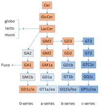
          Gangliosides constitute a subgroup of glycosphingolipids characterized by the presence of sialic acid residues in their structure. As constituents of cellular membranes, in particular of raft microdomains, they exert multiple functions, some of them capital in cell homeostasis. Their presence in cells is tightly regulated by a balanced expression and function of the enzymes responsible for their biosynthesis, ganglioside synthases, and their degradation, glycosidases. The dysregulation of their abundance results in rare and common diseases. In this review, we make a point on the relevance of gangliosides and some of their metabolic precursors, such as ceramides, in the function of podocytes, the main cellular component of the glomerular filtration barrier, as well as their implications in podocytopathies. The results presented in this review suggest the pertinence of clinical lipidomic studies targeting these metabolites.

          Related collections

          Most cited references84

          • Record: found
          • Abstract: found
          • Article: not found

          Revitalizing membrane rafts: new tools and insights.

          Ten years ago, we wrote a Review on lipid rafts and signalling in the launch issue of Nature Reviews Molecular Cell Biology. At the time, this field was suffering from ambiguous methodology and imprecise nomenclature. Now, new techniques are deepening our insight into the dynamics of membrane organization. Here, we discuss how the field has matured and present an evolving model in which membranes are occupied by fluctuating nanoscale assemblies of sphingolipids, cholesterol and proteins that can be stabilized into platforms that are important in signalling, viral infection and membrane trafficking.
            Bookmark
            • Record: found
            • Abstract: found
            • Article: not found

            Rituximab targets podocytes in recurrent focal segmental glomerulosclerosis.

            Focal segmental glomerulosclerosis (FSGS) is a glomerular disease characterized by proteinuria, progression to end-stage renal disease, and recurrence of proteinuria after kidney transplantation in about one-third of patients. It has been suggested that rituximab might treat recurrent FSGS through an unknown mechanism. Rituximab not only recognizes CD20 on B lymphocytes, but might also bind sphingomyelin phosphodiesterase acid-like 3b (SMPDL-3b) protein and regulate acid sphingomyelinase (ASMase) activity. We hypothesized that rituximab prevents recurrent FSGS and preserves podocyte SMPDL-3b expression. We studied 41 patients at high risk for recurrent FSGS, 27 of whom were treated with rituximab at time of kidney transplant. SMPDL-3b protein, ASMase activity, and cytoskeleton remodeling were studied in cultured normal human podocytes that had been exposed to patient sera with or without rituximab. Rituximab treatment was associated with lower incidence of posttransplant proteinuria and stabilization of glomerular filtration rate. The number of SMPDL-3b(+) podocytes in postreperfusion biopsies was reduced in patients who developed recurrent FSGS. Rituximab partially prevented SMPDL-3b and ASMase down-regulation that was observed in podocytes treated with the sera of patients with recurrent FSGS. Overexpression of SMPDL-3b or treatment with rituximab was able to prevent disruption of the actin cytoskeleton and podocyte apoptosis induced by patient sera. This effect was diminished in cultured podocytes where SMPDL-3b was silenced. Our study suggests that treatment of high-risk patients with rituximab at time of kidney transplant might prevent recurrent FSGS by modulating podocyte function in an SMPDL-3b-dependent manner.
              Bookmark
              • Record: found
              • Abstract: found
              • Article: not found

              Dissociation of the insulin receptor and caveolin-1 complex by ganglioside GM3 in the state of insulin resistance.

              Membrane microdomains (lipid rafts) are now recognized as critical for proper compartmentalization of insulin signaling. We previously demonstrated that, in adipocytes in a state of TNFalpha-induced insulin resistance, the inhibition of insulin metabolic signaling and the elimination of insulin receptors (IR) from the caveolae microdomains were associated with an accumulation of the ganglioside GM3. To gain insight into molecular mechanisms behind interactions of IR, caveolin-1 (Cav1), and GM3 in adipocytes, we have performed immunoprecipitations, cross-linking studies of IR and GM3, and live cell studies using total internal reflection fluorescence microscopy and fluorescence recovery after photobleaching techniques. We found that (i) IR form complexes with Cav1 and GM3 independently; (ii) in GM3-enriched membranes the mobility of IR is increased by dissociation of the IR-Cav1 interaction; and (iii) the lysine residue localized just above the transmembrane domain of the IR beta-subunit is essential for the interaction of IR with GM3. Because insulin metabolic signal transduction in adipocytes is known to be critically dependent on caveolae, we propose a pathological feature of insulin resistance in adipocytes caused by dissociation of the IR-Cav1 complex by the interactions of IR with GM3 in microdomains.
                Bookmark

                Author and article information

                Journal
                Int J Mol Sci
                Int J Mol Sci
                ijms
                International Journal of Molecular Sciences
                MDPI
                1422-0067
                17 December 2020
                December 2020
                : 21
                : 24
                : 9645
                Affiliations
                [1 ]INSERM, IMRB, Univ Paris Est Créteil, F-94010 Créteil, France; berkan.savas@ 123456inserm.fr (B.S.); dil.sahali@ 123456inserm.fr (D.S.)
                [2 ]Department of Biochemistry and Molecular & Cellular Biology, Georgetown University, 20007 Washington, DC, USA; gastarita@ 123456gmail.com
                [3 ]Department of Medical Biotechnology and Translational Medicine, University of Milano, Milano Italy, 20090 Segrate (Milano), Italy; massimo.aureli@ 123456unimi.it
                [4 ]Service Néphrologie, AP-HP, Hôpital Henri Mondor, F-94010 Créteil, France
                Author notes
                [* ]Correspondence: mario.ollero@ 123456inserm.fr ; Tel.: +33-149813667
                Author information
                https://orcid.org/0000-0001-6700-301X
                https://orcid.org/0000-0003-3590-706X
                Article
                ijms-21-09645
                10.3390/ijms21249645
                7766259
                33348903
                3518fb7f-ceef-4bc4-93b5-bb28cfc7f4e8
                © 2020 by the authors.

                Licensee MDPI, Basel, Switzerland. This article is an open access article distributed under the terms and conditions of the Creative Commons Attribution (CC BY) license ( http://creativecommons.org/licenses/by/4.0/).

                History
                : 23 November 2020
                : 14 December 2020
                Categories
                Review

                Molecular biology
                glycosphingolipids,ceramide,podocytopathies,glomerulopathies,glomerulus,kidney,rafts,nephrotic syndrome

                Comments

                Comment on this article