1
views
0
recommends
+1 Recommend
0 collections
    0
    shares
      • Record: found
      • Abstract: found
      • Article: found
      Is Open Access

      Diagnostic Potential of Plasma IgA1 O-Glycans in Discriminating IgA Nephropathy From Other Glomerular Diseases and Healthy Participants

      research-article

      Read this article at

      Bookmark
          There is no author summary for this article yet. Authors can add summaries to their articles on ScienceOpen to make them more accessible to a non-specialist audience.

          Abstract

          Background: Aberrant O-glycosylation of IgA1 plays an important role in IgA nephropathy pathogenesis. Previous proteomic studies analyzed O-glycans of the circulating IgA1 hinge region and found that the N-acetylgalactosamine (GalNAc) and galactose numbers in the hinge region of IgA1 of patients with IgA nephropathy were lower than those in healthy participants. However, the diagnostic performance of the O-glycosylation traits in the hinge region of plasma IgA1 for IgA nephropathy remains unelucidated. The present study aimed to determine the difference in plasma IgA1 hinge region O-glycoforms among IgA nephropathy, non-IgA nephropathy disease controls, and healthy participants, and to further evaluate the diagnostic performance of plasma IgA1 glycosylation traits.

          Methods: Sixty-two patients with biopsy-proven primary IgA nephropathy, 30 age- and sex-matched non-IgA nephropathy disease controls (10 patients with membranous nephropathy, 10 with focal segmental glomerulosclerosis, and 10 with minimal change disease), and 30 healthy participants were prospectively recruited. Plasma galactose deficient-IgA1 levels were measured using a KM55 kit. Plasma IgA was extracted using IgA immunoaffinity beads. After de-N-glycosylation, reduction, alkylation, trypsin digestion, and O-glycopeptide enrichment via hydrophilic interaction liquid chromatography, liquid chromatography tandem mass spectrometry (LC-MS/MS) was applied to analyze the IgA1 O-glycosylation patterns and we derived the plasma IgA1 O-glycosylation traits.

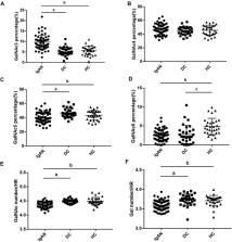
          Results: Plasma IgA1 O-glycosylation patterns were significantly changed in IgA nephropathy patients compared to those with non-IgA nephropathy disease controls and healthy participants. The GalNAc number was lowest in IgA nephropathy patients. In addition, a similar result was observed for the galactose number in the IgA1 hinge region. These values showed moderate potential for discriminating between IgA nephropathy and the controls. When these values were combined, the area under the curve increased compared to when they were considered individually. When further adding a clinical indicator, the area under the curve of the GalNAc-galactose-IgA panel exceed 0.9 in discriminating IgA nephropathy from the controls.

          Conclusion: The amount of GalNAc and galactose in plasma IgA1 hinge region identified by glycoproteomics could be used as a diagnostic biomarker for IgA nephropathy. The panel containing GalNAc, galactose, and circulating IgA displayed excellent diagnostic performance and is promising for practical clinical applications.

          Related collections

          Most cited references28

          • Record: found
          • Abstract: found
          • Article: not found

          Universal sample preparation method for proteome analysis.

          We describe a method, filter-aided sample preparation (FASP), which combines the advantages of in-gel and in-solution digestion for mass spectrometry-based proteomics. We completely solubilized the proteome in sodium dodecyl sulfate, which we then exchanged by urea on a standard filtration device. Peptides eluted after digestion on the filter were pure, allowing single-run analyses of organelles and an unprecedented depth of proteome coverage.
            Bookmark
            • Record: found
            • Abstract: found
            • Article: not found

            Oxford Classification of IgA nephropathy 2016: an update from the IgA Nephropathy Classification Working Group.

            Since the Oxford Classification of IgA nephropathy (IgAN) was published in 2009, MEST scores have been increasingly used in clinical practice. Further retrospective cohort studies have confirmed that in biopsy specimens with a minimum of 8 glomeruli, mesangial hypercellularity (M), segmental sclerosis (S), and interstitial fibrosis/tubular atrophy (T) lesions predict clinical outcome. In a larger, more broadly based cohort than in the original Oxford study, crescents (C) are predictive of outcome, and we now recommend that C be added to the MEST score, and biopsy reporting should provide a MEST-C score. Inconsistencies in the reporting of M and endocapillary cellularity (E) lesions have been reported, so a web-based educational tool to assist pathologists has been developed. A large study showed E lesions are predictive of outcome in children and adults, but only in those without immunosuppression. A review of S lesions suggests there may be clinical utility in the subclassification of segmental sclerosis, identifying those cases with evidence of podocyte damage. It has now been shown that combining the MEST score with clinical data at biopsy provides the same predictive power as monitoring clinical data for 2 years; this requires further evaluation to assess earlier effective treatment intervention. The IgAN Classification Working Group has established a well-characterized dataset from a large cohort of adults and children with IgAN that will provide a substrate for further studies to refine risk prediction and clinical utility, including the MEST-C score and other factors.
              Bookmark
              • Record: found
              • Abstract: not found
              • Article: not found

              Estimating GFR using the CKD Epidemiology Collaboration (CKD-EPI) creatinine equation: more accurate GFR estimates, lower CKD prevalence estimates, and better risk predictions.

                Bookmark

                Author and article information

                Contributors
                Journal
                Front Mol Biosci
                Front Mol Biosci
                Front. Mol. Biosci.
                Frontiers in Molecular Biosciences
                Frontiers Media S.A.
                2296-889X
                04 April 2022
                2022
                : 9
                : 871615
                Affiliations
                [1] 1 Department of Nephrology , State Key Laboratory of Complex Severe and Rare Diseases , Peking Union Medical College Hospital , Chinese Academy of Medical Science and Peking Union Medical College , Beijing, China
                [2] 2 Core Facility of Instruments , School of Basic Medicine , Peking Union Medical College , Institute of Basic Medical Sciences , Chinese Academy of Medical Sciences , Beijing, China
                [3] 3 Medical Research Center , State Key Laboratory of Complex Severe and Rare Diseases , Peking Union Medical College Hospital , Chinese Academy of Medical Science and Peking Union Medical College , Beijing, China
                [4] 4 State Key Laboratory of Bioactive Substances and Functions of Natural Medicines , Institute of Materia Medica , Peking Union Medical College , Chinese Academy of Medical Sciences , Beijing, China
                [5] 5 Department of Nephrology , The First Medical Centre , Chinese PLA General Hospital , Medical School of Chinese PLA , Beijing, China
                Author notes

                Edited by: Yang Zhao, National Institute of Metrology, China

                Reviewed by: Jicheng Lv, Peking University First Hospital, China

                Haihong Bai, Capital Medical University, China

                [ † ]

                These authors have contributed equally to this work

                This article was submitted to Glycoscience, a section of the journal Frontiers in Molecular Biosciences

                Article
                871615
                10.3389/fmolb.2022.871615
                9014244
                35445079
                3970f7ac-363c-44ed-baca-41598b866538
                Copyright © 2022 Zhang, Sun, Zhang, Li, Guo, Ye, Cai, Sun and Li.

                This is an open-access article distributed under the terms of the Creative Commons Attribution License (CC BY). The use, distribution or reproduction in other forums is permitted, provided the original author(s) and the copyright owner(s) are credited and that the original publication in this journal is cited, in accordance with accepted academic practice. No use, distribution or reproduction is permitted which does not comply with these terms.

                History
                : 08 February 2022
                : 21 March 2022
                Categories
                Molecular Biosciences
                Original Research

                iga nephropathy,o-glycosylation,iga1 hinge region,glycoproteomics,biomarker

                Comments

                Comment on this article