51
views
0
recommends
+1 Recommend
0 collections
    0
    shares
      • Record: found
      • Abstract: found
      • Article: found
      Is Open Access

      Heterogeneous induction of microglia M2a phenotype by central administration of interleukin-4

      research-article

      Read this article at

      Bookmark
          There is no author summary for this article yet. Authors can add summaries to their articles on ScienceOpen to make them more accessible to a non-specialist audience.

          Abstract

          Background

          Acquisition of the M1 or M2 phenotypes by microglia has been shown to occur during the development of pathological conditions, with M1 activation being widely involved in neurotoxicity in relation with the anatomical localization and the reactivity of subtypes of microglia cells. On the contrary, little is known on the ability of microglia to undergo M2 polarization by interleukin-4 (IL4), the typical M2a polarization signal for peripheral macrophages.

          Methods

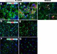
          Recombinant mouse IL4 was injected in the third cerebral ventricle of mice to induce brain alternative polarization. The mRNA levels of Fizz1, Arg1, and Ym1 genes, known to be up-regulated by IL4 in peripheral macrophages, together with additional polarization markers, were evaluated in the striatum and frontal cortex at different time intervals after central administration of IL4; in parallel, M2a protein expression was evaluated in tissue extracts and at the cellular level.

          Results

          Our results show that the potency and temporal profile of IL4-mediated M2a gene induction vary depending on the gene analyzed and according to the specific brain area analyzed, with the striatum showing a reduced M2a response compared with the frontal cortex, as further substantiated by assays of polarization protein levels. Of notice, Fizz1 mRNA induction reached 100-fold level, underscoring the potency of this specific IL4 signaling pathway in the brain. In addition, immunochemistry assays demonstrated the localization of the M2 response specifically to microglia cells and, more interestingly, the existence of a subpopulation of microglia cells amenable to undergoing M2a polarization in the healthy mouse brain.

          Conclusions

          These results show that the responsiveness of brain macrophages to centrally administered IL4 may vary depending on the gene and brain area analyzed, and that M2a polarization can be ascribed to a subpopulation of IL4-responsive microglia cells. The biochemical pathways that enable microglia to undergo M2a activation represent key aspects for understanding the physiopathology of neuroinflammation and for developing novel therapeutic and diagnostic agents.

          Electronic supplementary material

          The online version of this article (doi:10.1186/s12974-014-0211-6) contains supplementary material, which is available to authorized users.

          Related collections

          Most cited references26

          • Record: found
          • Abstract: found
          • Article: not found

          Systemic LPS causes chronic neuroinflammation and progressive neurodegeneration.

          Inflammation is implicated in the progressive nature of neurodegenerative diseases, such as Parkinson's disease, but the mechanisms are poorly understood. A single systemic lipopolysaccharide (LPS, 5 mg/kg, i.p.) or tumor necrosis factor alpha (TNFalpha, 0.25 mg/kg, i.p.) injection was administered in adult wild-type mice and in mice lacking TNFalpha receptors (TNF R1/R2(-/-)) to discern the mechanisms of inflammation transfer from the periphery to the brain and the neurodegenerative consequences. Systemic LPS administration resulted in rapid brain TNFalpha increase that remained elevated for 10 months, while peripheral TNFalpha (serum and liver) had subsided by 9 h (serum) and 1 week (liver). Systemic TNFalpha and LPS administration activated microglia and increased expression of brain pro-inflammatory factors (i.e., TNFalpha, MCP-1, IL-1beta, and NF-kappaB p65) in wild-type mice, but not in TNF R1/R2(-/-) mice. Further, LPS reduced the number of tyrosine hydroxylase-immunoreactive neurons in the substantia nigra (SN) by 23% at 7-months post-treatment, which progressed to 47% at 10 months. Together, these data demonstrate that through TNFalpha, peripheral inflammation in adult animals can: (1) activate brain microglia to produce chronically elevated pro-inflammatory factors; (2) induce delayed and progressive loss of DA neurons in the SN. These findings provide valuable insight into the potential pathogenesis and self-propelling nature of Parkinson's disease. (c) 2007 Wiley-Liss, Inc.
            Bookmark
            • Record: found
            • Abstract: found
            • Article: not found

            Heterogeneity of Microglial Activation in the Innate Immune Response in the Brain

            The immune response in the brain has been widely investigated and while many studies have focused on the proinflammatory cytotoxic response, the brain’s innate immune system demonstrates significant heterogeneity. Microglia, like other tissue macrophages, participate in repair and resolution processes after infection or injury to restore normal tissue homeostasis. This review examines the mechanisms that lead to reduction of self-toxicity and to repair and restructuring of the damaged extracellular matrix in the brain. Part of the resolution process involves switching macrophage functional activation to include reduction of proinflammatory mediators, increased production and release of anti-inflammatory cytokines, and production of cytoactive factors involved in repair and reconstruction of the damaged brain. Two partially overlapping and complimentary functional macrophage states have been identified and are called alternative activation and acquired deactivation. The immunosuppressive and repair processes of each of these states and how alternative activation and acquired deactivation participate in chronic neuroinflammation in the brain are discussed.
              Bookmark
              • Record: found
              • Abstract: found
              • Article: not found

              Inflammation as a causative factor in the aetiology of Parkinson's disease.

              Parkinson's disease (PD) is a progressive neurodegenerative disorder affecting mainly the elderly, although a small proportion of PD patients develop the illness at a much younger age. In the former group, idiopathic PD patients, the causes of the illness have been the subject of longstanding debate with environmental toxins, mitochondrial dysfunction, abnormal protein handling and oxidative stress being suggested. One problem has been that the epidemiology of PD has offered few clues to provide evidence for a single major causative factor. Comparatively recently it has been found that in both patients and experimental models of PD in animals neuroinflammation appears to be a ubiquitous finding. These cases present with all of the classical features of inflammation including phagocyte activation, increased synthesis and release of proinflammatory cytokines and complement activation. Although this process is vital for normal function and protection in both the CNS, as in the periphery, it is postulated that in the aetiology of PD this process may spiral out of control with over activation of microglia, over production of cytokines and other proinflammatory mediators as well as the release of destructive molecules such as reactive oxygen species. Given that dopaminergic neurons in the substantia nigra are relatively vulnerable to 'stress' and the region has a large population of microglia in comparison to other CNS structures, these events may easily trigger neurodegeneration. These factors are examined in this review along with a consideration of the possible use of anti-inflammatory drugs in PD.
                Bookmark

                Author and article information

                Contributors
                giovanna.pepe@unimi.it
                giorgia.calderazzi@unimi.it
                marcella.demaglie@unimi.it
                alessandromaria.villa@unimi.it
                elisabetta.vegeto@unimi.it
                Journal
                J Neuroinflammation
                J Neuroinflammation
                Journal of Neuroinflammation
                BioMed Central (London )
                1742-2094
                31 December 2014
                31 December 2014
                2014
                : 11
                : 1
                : 211
                Affiliations
                [ ]Center of Excellence on Neurodegenerative Diseases, Department of Pharmacological and Biomolecular Sciences, University of Milan, via Balzaretti, 9, 20133 Milan, Italy
                [ ]Department of Veterinary Science and Public Health Veterinary Medicine, University of Milan, Via Celoria, 20133 Milan, Italy
                Article
                211
                10.1186/s12974-014-0211-6
                4302702
                25551794
                39852be6-460c-40c4-912c-6346dc4f3d89
                © Pepe et al.; licensee BioMed Central. 2014

                This is an Open Access article distributed under the terms of the Creative Commons Attribution License ( http://creativecommons.org/licenses/by/4.0), which permits unrestricted use, distribution, and reproduction in any medium, provided the original work is properly credited. The Creative Commons Public Domain Dedication waiver ( http://creativecommons.org/publicdomain/zero/1.0/) applies to the data made available in this article, unless otherwise stated.

                History
                : 24 July 2014
                : 2 December 2014
                Categories
                Research
                Custom metadata
                © The Author(s) 2014

                Neurosciences
                alternative activation,centrally administered interleukin-4,m2 polarization,microglia

                Comments

                Comment on this article