Inviting an author to review:
Find an author and click ‘Invite to review selected article’ near their name.
Search for authorsSearch for similar articles
9
views
0
recommends
+1 Recommend
0 collections
    0
    shares
      • Record: found
      • Abstract: found
      • Article: found
      Is Open Access

      Extracellular Vesicles as Mediators of Cellular Crosstalk Between Immune System and Kidney Graft

      review-article

      Read this article at

      Bookmark
          There is no author summary for this article yet. Authors can add summaries to their articles on ScienceOpen to make them more accessible to a non-specialist audience.

          Abstract

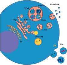
          Extracellular vesicles (EVs) are known immune-modulators exerting a critical role in kidney transplantation (KT). EV bioactive cargo includes graft antigens, costimulatory/inhibitory molecules, cytokines, growth factors, and functional microRNAs (miRNAs) that may modulate expression of recipient cell genes. As paracrine factors, neutrophil- and macrophage-derived EVs exert immunosuppressive and immune-stimulating effects on dendritic cells, respectively. Dendritic cell-derived EVs mediate alloantigen spreading and modulate antigen presentation to T lymphocytes. At systemic level, EVs exert pleiotropic effects on complement and coagulation. Depending on their biogenesis, they can amplify complement activation or shed complement inhibitors and prevent cell lysis. Likewise, endothelial- and platelet-derived EVs can exert procoagulant/prothrombotic effects and also promote endothelial survival and angiogenesis after ischemic injury. Kidney endothelial- and tubular-derived EVs play a key role in ischemia–reperfusion injury (IRI) and during the healing process; additionally, they can trigger rejection by inducing both alloimmune and autoimmune responses. Endothelial EVs have procoagulant/pro-inflammatory effects and can release sequestered self-antigens, generating a tissue-specific autoimmunity. Renal tubule-derived EVs shuttle pro-fibrotic mediators (TGF-β and miR-21) to interstitial fibroblasts and modulate neutrophil and T-lymphocyte influx. These processes can lead to peritubular capillary rarefaction and interstitial fibrosis–tubular atrophy. Different EVs, including those from mesenchymal stromal cells (MSCs), have been employed as a therapeutic tool in experimental models of rejection and IRI. These particles protect tubular and endothelial cells (by inhibition of apoptosis and inflammation–fibrogenesis or by inducing autophagy) and stimulate tissue regeneration (by triggering angiogenesis, cell proliferation, and migration). Finally, urinary and serum EVs represent potential biomarkers for delayed graft function (DGF) and acute rejection. In conclusion, EVs sustain an intricate crosstalk between graft tissue and innate/adaptive immune systems. EVs play a major role in allorecognition, IRI, autoimmunity, and alloimmunity and are promising as biomarkers and therapeutic tools in KT.

          Related collections

          Most cited references179

          • Record: found
          • Abstract: found
          • Article: not found

          Mechanisms of maladaptive repair after AKI leading to accelerated kidney ageing and CKD.

          Acute kidney injury is an increasingly common complication of hospital admission and is associated with high levels of morbidity and mortality. A hypotensive, septic, or toxic insult can initiate a cascade of events, resulting in impaired microcirculation, activation of inflammatory pathways and tubular cell injury or death. These processes ultimately result in acutely impaired kidney function and initiation of a repair response. This Review explores the various mechanisms responsible for the initiation and propagation of acute kidney injury, the prototypic mechanisms by which a substantially damaged kidney can regenerate its normal architecture, and how the adaptive processes of repair can become maladaptive. These mechanisms, which include G2/M cell-cycle arrest, cell senescence, profibrogenic cytokine production, and activation of pericytes and interstitial myofibroblasts, contribute to the development of progressive fibrotic kidney disease. The end result is a state that mimics accelerated kidney ageing. These mechanisms present important opportunities for the design of targeted therapeutic strategies to promote adaptive renal recovery and minimize progressive fibrosis and chronic kidney disease after acute insults.
            Bookmark
            • Record: found
            • Abstract: found
            • Article: found
            Is Open Access

            Apoptotic Cell-Derived Extracellular Vesicles: More Than Just Debris

            The many functions of extracellular vesicles (EVs) like exosomes and microvesicles released from healthy cells have been well characterized, particularly in relation to their roles in immune modulation. Apoptotic bodies, a major class of EV released as a product of apoptotic cell disassembly, and other types of EVs released from dying cells are also becoming recognized as key players in this emerging field. There is now increasing evidence to suggest that EVs produced during apoptosis have important immune regulatory roles, a concept relevant across different disease settings including autoimmunity, cancer, and infection. Therefore, this review focuses on how the formation of EVs during apoptosis could be a key mechanism of immune modulation by dying cells.
              Bookmark
              • Record: found
              • Abstract: found
              • Article: not found

              Ischemia/Reperfusion.

              Ischemic disorders, such as myocardial infarction, stroke, and peripheral vascular disease, are the most common causes of debilitating disease and death in westernized cultures. The extent of tissue injury relates directly to the extent of blood flow reduction and to the length of the ischemic period, which influence the levels to which cellular ATP and intracellular pH are reduced. By impairing ATPase-dependent ion transport, ischemia causes intracellular and mitochondrial calcium levels to increase (calcium overload). Cell volume regulatory mechanisms are also disrupted by the lack of ATP, which can induce lysis of organelle and plasma membranes. Reperfusion, although required to salvage oxygen-starved tissues, produces paradoxical tissue responses that fuel the production of reactive oxygen species (oxygen paradox), sequestration of proinflammatory immunocytes in ischemic tissues, endoplasmic reticulum stress, and development of postischemic capillary no-reflow, which amplify tissue injury. These pathologic events culminate in opening of mitochondrial permeability transition pores as a common end-effector of ischemia/reperfusion (I/R)-induced cell lysis and death. Emerging concepts include the influence of the intestinal microbiome, fetal programming, epigenetic changes, and microparticles in the pathogenesis of I/R. The overall goal of this review is to describe these and other mechanisms that contribute to I/R injury. Because so many different deleterious events participate in I/R, it is clear that therapeutic approaches will be effective only when multiple pathologic processes are targeted. In addition, the translational significance of I/R research will be enhanced by much wider use of animal models that incorporate the complicating effects of risk factors for cardiovascular disease. © 2017 American Physiological Society. Compr Physiol 7:113-170, 2017.
                Bookmark

                Author and article information

                Contributors
                Journal
                Front Immunol
                Front Immunol
                Front. Immunol.
                Frontiers in Immunology
                Frontiers Media S.A.
                1664-3224
                27 February 2020
                2020
                : 11
                : 74
                Affiliations
                [1] 1Nephrology and Kidney Transplantation Unit, Department of Translational Medicine, University of Piemonte Orientale (UPO) , Novara, Italy
                [2] 2Center for Autoimmune and Allergic Diseases (CAAD), University of Piemonte Orientale (UPO) , Novara, Italy
                [3] 3Division of Hematology/Medical Oncology, Icahn School of Medicine at Mount Sinai Hospital, The Tisch Cancer Institute , New York, NY, United States
                [4] 4Nephrology Dialysis and Transplantation Unit, Department of Medical and Surgical Sciences, University of Foggia , Foggia, Italy
                Author notes

                Edited by: Guseppe Grandaliano, University of Foggia, Italy

                Reviewed by: Xiangyang Zhu, Mayo Clinic, United States; Francesco Paolo Schena, University of Bari Aldo Moro, Italy

                *Correspondence: Vincenzo Cantaluppi vincenzo.cantaluppi@ 123456med.uniupo.it

                This article was submitted to Molecular Innate Immunity, a section of the journal Frontiers in Immunology

                Article
                10.3389/fimmu.2020.00074
                7057849
                32180768
                3bf019c1-a38c-4088-8a4a-f2c8eb65ed16
                Copyright © 2020 Quaglia, Dellepiane, Guglielmetti, Merlotti, Castellano and Cantaluppi.

                This is an open-access article distributed under the terms of the Creative Commons Attribution License (CC BY). The use, distribution or reproduction in other forums is permitted, provided the original author(s) and the copyright owner(s) are credited and that the original publication in this journal is cited, in accordance with accepted academic practice. No use, distribution or reproduction is permitted which does not comply with these terms.

                History
                : 06 November 2019
                : 13 January 2020
                Page count
                Figures: 3, Tables: 4, Equations: 0, References: 239, Pages: 19, Words: 14982
                Categories
                Immunology
                Review

                Immunology
                extracellular vesicles,acute rejection,ischemia-reperfusion syndrome,autoimmunity,renal transplant,biomarker,mirna

                Comments

                Comment on this article