11
views
0
recommends
+1 Recommend
0 collections
    0
    shares
      • Record: found
      • Abstract: found
      • Article: found
      Is Open Access

      Cytotoxic lanthanum oxide nanoparticles sensitize glioblastoma cells to radiation therapy and temozolomide: an in vitro rationale for translational studies

      research-article
      1 , 2 , , 1 , 1
      Scientific Reports
      Nature Publishing Group UK
      CNS cancer, Nanoparticles, Cell death

      Read this article at

      Bookmark
          There is no author summary for this article yet. Authors can add summaries to their articles on ScienceOpen to make them more accessible to a non-specialist audience.

          Abstract

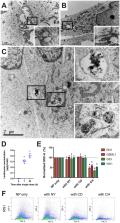
          Glioblastoma (GBM) is a malignant brain tumour with a dismal prognosis, despite best treatment by surgical resection, radiation therapy (RT) and chemotherapy with temozolomide (TMZ). Nanoparticle (NP) therapy is an emerging consideration due to the ability of NPs to be formulated and cross the blood brain barrier. Lanthanum oxide (La 2O 3) NPs are therapeutically advantageous due to the unique chemical properties of lanthanum making it cytotoxic to cancers, and able to enhance existing anti-cancer treatments. However, La 2O 3 NPs have yet to be thoroughly investigated in brain tumors. We show that these NPs can reach the brain after venous injection, penetrate into GBM cells via endocytosis, dissociate to be cytotoxic, and enhance the therapeutic effects of RT and TMZ. The mechanisms of cell death by La 2O 3 NPs were found to be multifaceted. Increasing NP concentration was correlated to increased intrinsic and extrinsic apoptosis pathway markers in a radical oxygen species (ROS)-dependent manner, as well as involving direct DNA damage and autophagic pathways within GBM patient-derived cell lines. NP interactions to sensitize GBM to RT and TMZ were shown to involve these pathways by enhancing ROS and apoptotic mechanisms. We therefore demonstrate the therapeutic potential of La 2O 3 NPs to treat GBM cells in vitro, and encourage translational exploration in the future.

          Related collections

          Most cited references60

          • Record: found
          • Abstract: found
          • Article: not found

          Radiotherapy plus Concomitant and Adjuvant Temozolomide for Glioblastoma

          Glioblastoma, the most common primary brain tumor in adults, is usually rapidly fatal. The current standard of care for newly diagnosed glioblastoma is surgical resection to the extent feasible, followed by adjuvant radiotherapy. In this trial we compared radiotherapy alone with radiotherapy plus temozolomide, given concomitantly with and after radiotherapy, in terms of efficacy and safety. Patients with newly diagnosed, histologically confirmed glioblastoma were randomly assigned to receive radiotherapy alone (fractionated focal irradiation in daily fractions of 2 Gy given 5 days per week for 6 weeks, for a total of 60 Gy) or radiotherapy plus continuous daily temozolomide (75 mg per square meter of body-surface area per day, 7 days per week from the first to the last day of radiotherapy), followed by six cycles of adjuvant temozolomide (150 to 200 mg per square meter for 5 days during each 28-day cycle). The primary end point was overall survival. A total of 573 patients from 85 centers underwent randomization. The median age was 56 years, and 84 percent of patients had undergone debulking surgery. At a median follow-up of 28 months, the median survival was 14.6 months with radiotherapy plus temozolomide and 12.1 months with radiotherapy alone. The unadjusted hazard ratio for death in the radiotherapy-plus-temozolomide group was 0.63 (95 percent confidence interval, 0.52 to 0.75; P<0.001 by the log-rank test). The two-year survival rate was 26.5 percent with radiotherapy plus temozolomide and 10.4 percent with radiotherapy alone. Concomitant treatment with radiotherapy plus temozolomide resulted in grade 3 or 4 hematologic toxic effects in 7 percent of patients. The addition of temozolomide to radiotherapy for newly diagnosed glioblastoma resulted in a clinically meaningful and statistically significant survival benefit with minimal additional toxicity. Copyright 2005 Massachusetts Medical Society.
            Bookmark
            • Record: found
            • Abstract: found
            • Article: not found

            Role of reactive oxygen species (ROS) in apoptosis induction.

            Reactive oxygen species (ROS) and mitochondria play an important role in apoptosis induction under both physiologic and pathologic conditions. Interestingly, mitochondria are both source and target of ROS. Cytochrome c release from mitochondria, that triggers caspase activation, appears to be largely mediated by direct or indirect ROS action. On the other hand, ROS have also anti-apoptotic effects. This review focuses on the role of ROS in the regulation of apoptosis, especially in inflammatory cells.
              Bookmark
              • Record: found
              • Abstract: not found
              • Article: not found

              Quantitative analysis of dose-effect relationships: the combined effects of multiple drugs or enzyme inhibitors

                Bookmark

                Author and article information

                Contributors
                victor.lu@jhsmiami.org
                Journal
                Sci Rep
                Sci Rep
                Scientific Reports
                Nature Publishing Group UK (London )
                2045-2322
                23 October 2020
                23 October 2020
                2020
                : 10
                : 18156
                Affiliations
                [1 ]GRID grid.1005.4, ISNI 0000 0004 4902 0432, Lowy Cancer Center, , University of New South Wales, ; Sydney, NSW Australia
                [2 ]GRID grid.26790.3a, ISNI 0000 0004 1936 8606, Department of Neurological Surgery, , University of Miami Miller School of Medicine, ; 1600 NW 10th Ave #1140, Miami, FL 33136 USA
                Article
                75372
                10.1038/s41598-020-75372-3
                7584621
                33097778
                3bf9fc56-696f-487d-a197-8f498004752f
                © The Author(s) 2020

                Open Access This article is licensed under a Creative Commons Attribution 4.0 International License, which permits use, sharing, adaptation, distribution and reproduction in any medium or format, as long as you give appropriate credit to the original author(s) and the source, provide a link to the Creative Commons licence, and indicate if changes were made. The images or other third party material in this article are included in the article's Creative Commons licence, unless indicated otherwise in a credit line to the material. If material is not included in the article's Creative Commons licence and your intended use is not permitted by statutory regulation or exceeds the permitted use, you will need to obtain permission directly from the copyright holder. To view a copy of this licence, visit http://creativecommons.org/licenses/by/4.0/.

                History
                : 12 March 2020
                : 14 October 2020
                Categories
                Article
                Custom metadata
                © The Author(s) 2020

                Uncategorized
                cns cancer,nanoparticles,cell death
                Uncategorized
                cns cancer, nanoparticles, cell death

                Comments

                Comment on this article