9
views
0
recommends
+1 Recommend
0 collections
    0
    shares
      • Record: found
      • Abstract: found
      • Article: not found

      Direct Ca 2+-dependent Heterophilic Interaction between Desmosomal Cadherins, Desmoglein and Desmocollin, Contributes to Cell–Cell Adhesion

      research-article
      ,
      The Journal of Cell Biology
      The Rockefeller University Press

      Read this article at

      ScienceOpenPublisherPMC
      Bookmark
          There is no author summary for this article yet. Authors can add summaries to their articles on ScienceOpen to make them more accessible to a non-specialist audience.

          Abstract

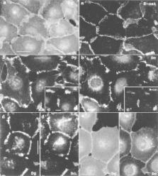
          Human fibrosarcoma cells, HT-1080, feature extensive adherens junctions, lack mature desmosomes, and express a single known desmosomal protein, Desmoglein 2 (Dsg2). Transfection of these cells with bovine Desmocollin 1a (Dsc1a) caused dramatic changes in the subcellular distribution of endogenous Dsg2. Both cadherins clustered in the areas of the adherens junctions, whereas only a minor portion of Dsg2 was seen in these areas in the parental cells. Deletion mapping showed that intact extracellular cadherin-like repeats of Dsc1a (Arg 1-Thr 170) are required for the translocation of Dsg2. Deletion of the intracellular C-domain that mediates the interaction of Dsc1a with plakoglobin, or the CSI region that is involved in the binding to desmoplakin, had no effect. Coimmunoprecipitation experiments of cell lysates stably expressing Dsc1a with anti-Dsc or -Dsg antibodies demonstrate that the desmosomal cadherins, Dsg2 and Dsc1a, are involved in a direct Ca 2+-dependent interaction. This conclusion was further supported by the results of solid phase binding experiments. These showed that the Dsc1a fragment containing cadherin-like repeats 1 and 2 binds directly to the extracellular portion of Dsg in a Ca 2+-dependent manner. The contribution of the Dsg/ Dsc interaction to cell–cell adhesion was tested by coculturing HT-1080 cells expressing Dsc1a with HT-1080 cells lacking Dsc but expressing myc-tagged plakoglobin (MPg). In the latter cells, MPg and the endogenous Dsg form stable complexes. The observed specific coimmunoprecipitation of MPg by anti-Dsc antibodies in coculture indicates that an intercellular interaction between Dsc1 and Dsg is involved in cell–cell adhesion.

          Related collections

          Most cited references42

          • Record: found
          • Abstract: found
          • Article: not found

          Cell adhesion: the molecular basis of tissue architecture and morphogenesis.

          A variety of cell adhesion mechanisms underlie the way that cells are organized in tissues. Stable cell interactions are needed to maintain the structural integrity of tissues, and dynamic changes in cell adhesion participate in the morphogenesis of developing tissues. Stable interactions actually require active adhesion mechanisms that are very similar to those involved in tissue dynamics. Adhesion mechanisms are highly regulated during tissue morphogenesis and are intimately related to the processes of cell motility and cell migration. In particular, the cadherins and the integrins have been implicated in the control of cell movement. Cadherin mediated cell compaction and cellular rearrangements may be analogous to integrin-mediated cell spreading and motility on the ECM. Regulation of cell adhesion can occur at several levels, including affinity modulation, clustering, and coordinated interactions with the actin cytoskeleton. Structural studies have begun to provide a picture of how the binding properties of adhesion receptors themselves might be regulated. However, regulation of tissue morphogenesis requires complex interactions between the adhesion receptors, the cytoskeleton, and networks of signaling pathways. Signals generated locally by the adhesion receptors themselves are involved in the regulation of cell adhesion. These regulatory pathways are also influenced by extrinsic signals arising from the classic growth factor receptors. Furthermore, signals generated locally be adhesion junctions can interact with classic signal transduction pathways to help control cell growth and differentiation. This coupling between physical adhesion and developmental signaling provides a mechanism to tightly integrate physical aspects of tissue morphogenesis with cell growth and differentiation, a coordination that is essential to achieve the intricate patterns of cells in tissues.
            Bookmark
            • Record: found
            • Abstract: found
            • Article: not found

            Structural basis of cell-cell adhesion by cadherins.

            Crystal structures of the amino-terminal domain of N-cadherin provide a picture at the atomic level of a specific adhesive contact between cells. A repeated set of dimer interfaces is common to the structure in three lattices. These interactions combine to form a linear zipper of molecules that mirrors the linear structure of the intracellular filaments with which cadherins associate. This cell-adhesion zipper may provide a mechanism to marshal individual molecular adhesive interactions into strong bonds between cells.
              Bookmark
              • Record: found
              • Abstract: found
              • Article: not found

              The cytoplasmic domain of the cell adhesion molecule uvomorulin associates with three independent proteins structurally related in different species.

              Uvomorulin belongs to the group of Ca2+-dependent cell adhesion molecules, which are integral membrane proteins with several structural features in common. In particular, the cytoplasmic part of these proteins is highly conserved in different species, suggesting a common biological function. To test this assumption we transfected a uvomorulin full-length cDNA into uvomorulin-negative mouse NIH 3T3 and L cells. Immunoprecipitations with anti-uvomorulin antibodies detected, in addition to uvomorulin, three independent proteins of 102, 88 and 80 kd which are of host origin and which form complexes with uvomorulin. Using cDNA constructs coding for uvomorulin with cytoplasmic or extracellular deletions it is shown that the 102, 88 and 80 kd proteins complex with the cytoplasmic domain of uvomorulin. Peptide pattern analysis revealed that these three proteins are identical in different mouse cells. When uvomorulin cDNA was introduced into cell lines from other species, such as human HeLa and avian fibroblasts, the expressed uvomorulin was also associated with endogenous 102, 88 and 80 kd proteins and, moreover, each of these proteins showed structural similarities to the respective mouse molecule. A panel of antibodies specific for known cytoplasmic proteins of mol. wts similar to those of the three proteins did not react with any of the described components. This suggests that the 102, 88 and 80 kd proteins constitute a new group of proteins for which we propose the nomenclature of catenin alpha, beta and gamma respectively. The characterization of these proteins provides a first molecular basis for a possible cytoplasmic anchorage of uvomorulin to the cytoskeleton.
                Bookmark

                Author and article information

                Journal
                J Cell Biol
                The Journal of Cell Biology
                The Rockefeller University Press
                0021-9525
                1540-8140
                14 July 1997
                : 138
                : 1
                : 193-201
                Affiliations
                Department of Dermatology, Washington University Medical School, St. Louis, Missouri 63110
                Article
                10.1083/jcb.138.1.193
                2139935
                9214392
                42e49a84-ac80-41b1-afac-7c950b2bc3e7
                Copyright @ 1997
                History
                : 7 January 1997
                : 1 May 1997
                Categories
                Article

                Cell biology
                Cell biology

                Comments

                Comment on this article