14
views
0
recommends
+1 Recommend
0 collections
    0
    shares
      • Record: found
      • Abstract: found
      • Article: found
      Is Open Access

      Perispinal Delivery of CNS Drugs

      research-article
      CNS Drugs
      Springer International Publishing

      Read this article at

      Bookmark
          There is no author summary for this article yet. Authors can add summaries to their articles on ScienceOpen to make them more accessible to a non-specialist audience.

          Abstract

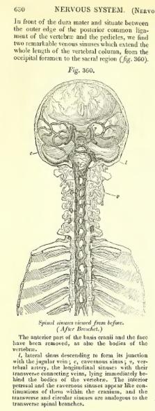
          Perispinal injection is a novel emerging method of drug delivery to the central nervous system (CNS). Physiological barriers prevent macromolecules from efficiently penetrating into the CNS after systemic administration. Perispinal injection is designed to use the cerebrospinal venous system (CSVS) to enhance delivery of drugs to the CNS. It delivers a substance into the anatomic area posterior to the ligamentum flavum, an anatomic region drained by the external vertebral venous plexus (EVVP), a division of the CSVS. Blood within the EVVP communicates with the deeper venous plexuses of the CSVS. The anatomical basis for this method originates in the detailed studies of the CSVS published in 1819 by the French anatomist Gilbert Breschet. By the turn of the century, Breschet’s findings were nearly forgotten, until rediscovered by American anatomist Oscar Batson in 1940. Batson confirmed the unique, linear, bidirectional and retrograde flow of blood between the spinal and cerebral divisions of the CSVS, made possible by the absence of venous valves. Recently, additional supporting evidence was discovered in the publications of American neurologist Corning. Analysis suggests that Corning’s famous first use of cocaine for spinal anesthesia in 1885 was in fact based on Breschet’s anatomical findings, and accomplished by perispinal injection. The therapeutic potential of perispinal injection for CNS disorders is highlighted by the rapid neurological improvement in patients with otherwise intractable neuroinflammatory disorders that may ensue following perispinal etanercept administration. Perispinal delivery merits intense investigation as a new method of enhanced delivery of macromolecules to the CNS and related structures.

          Electronic supplementary material

          The online version of this article (doi:10.1007/s40263-016-0339-2) contains supplementary material, which is available to authorized users.

          Related collections

          Most cited references107

          • Record: found
          • Abstract: not found
          • Article: not found

          THE FUNCTION OF THE VERTEBRAL VEINS AND THEIR ROLE IN THE SPREAD OF METASTASES.

          O V BATSON (1940)
            Bookmark
            • Record: found
            • Abstract: not found
            • Article: not found

            Intrathecal and epidural administration of opioids.

              Bookmark
              • Record: found
              • Abstract: found
              • Article: found
              Is Open Access

              Etanercept, a Widely Used Inhibitor of Tumor Necrosis Factor-α (TNF- α), Prevents Retinal Ganglion Cell Loss in a Rat Model of Glaucoma

              Background Visual loss in glaucoma is associated with pathological changes in retinal ganglion cell (RGC) axons and a slow decline in the RGC population. Age and elevated intraocular pressure (IOP) are the main risk factors for glaucomatous loss of vision. Several studies have implicated the proinflammatory cytokine tumor necrosis factor- α (TNF-α) as a link between elevated IOP and RGC death, but the cellular source of TNF-α and its causative role in RGC death remain uncertain. Here, using a rat model of glaucoma, we investigated the source of elevated TNF- α and examined whether Etanercept, a TNF-α blocker that is in common clinical use for other indications, is protective against RGC death. Methodology/Principal Findings Episcleral vein cauterization (EVC) caused intraocular pressure (IOP) to be elevated for at least 28 days. IOP elevation resulted in a dramatic increase in TNF-α levels within a few days, axonal degeneration, and a 38% loss of RGCs by 4 weeks. Immunostaining coupled with confocal microscopy showed that OHT induced robust induction of TNF-α in Iba-1-positive microglia around the optic nerve head (ONH). Despite persistent elevation of IOP, Etanercept reduced microglial activation, TNF-α levels, axon degeneration in the optic nerve, and the loss of RGCs. Conclusions/Significance Ocular hypertension (OHT) triggers an inflammatory response characterized by the appearance of activated microglia around the ONH that express TNF-α. Blocking TNF-α activity with a clinically approved agent inhibits this microglial response and prevents axonal degeneration and loss of RGCs. These findings suggest a new treatment strategy for glaucoma using TNF- α antagonists or suppressors of inflammation.
                Bookmark

                Author and article information

                Contributors
                (310) 479-0107 , nrimed@gmail.com
                Journal
                CNS Drugs
                CNS Drugs
                CNS Drugs
                Springer International Publishing (Cham )
                1172-7047
                27 April 2016
                27 April 2016
                2016
                : 30
                : 469-480
                Affiliations
                Institute of Neurological Recovery, 2300 Glades Road, Suite 305E, Boca Raton, FL 33431 USA
                Author information
                http://orcid.org/0000-0001-7614-499X
                Article
                339
                10.1007/s40263-016-0339-2
                4920856
                27120182
                48cd7ace-6895-4fb0-b64b-b04eddba0a8f
                © The Author(s) 2016

                Open AccessThis article is distributed under the terms of the Creative Commons Attribution-NonCommercial 4.0 International License (http://creativecommons.org/licenses/by-nc/4.0/), which permits any noncommercial use, distribution, and reproduction in any medium, provided you give appropriate credit to the original author(s) and the source, provide a link to the Creative Commons license, and indicate if changes were made.

                History
                Funding
                Funded by: Institute of Neurological Recovery
                Categories
                Leading Article
                Custom metadata
                © Springer International Publishing Switzerland 2016

                Comments

                Comment on this article