1
views
0
recommends
+1 Recommend
0 collections
    0
    shares
      • Record: found
      • Abstract: found
      • Article: found
      Is Open Access

      Label-free quantitative proteomics reveals the mechanisms of Aurora kinase B in renal cell carcinoma

      research-article

      Read this article at

      Bookmark
          There is no author summary for this article yet. Authors can add summaries to their articles on ScienceOpen to make them more accessible to a non-specialist audience.

          Abstract

          Background:

          Renal cell carcinoma is the most common form of kidney cancer which is a global threat to human health, needing to explore effective therapeutic targets and treatment methods. Aurora kinase B acts as an important carcinogenic role in various kinds of tumors, while its mechanism in renal cell carcinoma is indistinct. Herein we explore the underlying mechanism of Aurora kinase B in renal cell carcinoma.

          Methods and results:

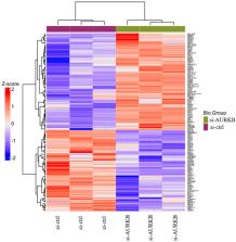
          Label-free quantitative proteomics analysis was employed to analyze the differentially expressed proteins in 786-O cells which were treated with si-Aurora kinase B or si-ctrl. In the current study, 169 differentially expressed proteins were identified. The top 10 upregulated proteins were MX2, IFI44L, ISG20, DDX58, F3, IFI44, ECE1, PRIC285, NIT1, and IFIT2. The top 10 downregulated proteins were FKBP9, FSTL1, DDAH1, TGFB2, HMGN3, COIL, FAM65A, PTPN14, ARFGAP2, and EIF2C2. GO enrichment analysis showed that these differentially expressed proteins participated in biological processes, including defense response to virus, response to virus, and type I interferon signaling pathway. These differentially expressed proteins participated in cellular components, including focal adhesion, cell-substrate adherens junction, cell-substrate junction, and endoplasmic reticulum lumen. These differentially expressed proteins participated in molecule functions, including guanyl nucleotide binding, nucleotidase activity, double-stranded RNA binding, 2′-5′-oligoadenylate synthetase activity, and virus receptor activity. Kyoto Encyclopedia of Genes and Genomes pathway analysis showed that the significantly changed proteins including OAS3, OAS2, JAK1, TAP1, and RAC1 were involved in Epstein-Barr virus infection.

          Conclusions:

          Taken together, our results demonstrate the possible mechanisms that Aurora kinase B may participate in renal cell carcinoma. These findings may provide insights into tumorigenesis and a theoretical basis for developing potential therapies of renal cell carcinoma.

          Related collections

          Most cited references41

          • Record: found
          • Abstract: found
          • Article: not found

          Renal cell carcinoma

          Renal cell carcinoma (RCC) denotes cancer originated from the renal epithelium and accounts for >90% of cancers in the kidney. The disease encompasses >10 histological and molecular subtypes, of which clear cell RCC (ccRCC) is most common and accounts for most cancer-related deaths. Although somatic VHL mutations have been described for some time, more-recent cancer genomic studies have identified mutations in epigenetic regulatory genes and demonstrated marked intra-tumour heterogeneity, which could have prognostic, predictive and therapeutic relevance. Localized RCC can be successfully managed with surgery, whereas metastatic RCC is refractory to conventional chemotherapy. However, over the past decade, marked advances in the treatment of metastatic RCC have been made, with targeted agents including sorafenib, sunitinib, bevacizumab, pazopanib and axitinib, which inhibit vascular endothelial growth factor (VEGF) and its receptor (VEGFR), and everolimus and temsirolimus, which inhibit mechanistic target of rapamycin complex 1 (mTORC1), being approved. Since 2015, agents with additional targets aside from VEGFR have been approved, such as cabozantinib and lenvatinib; immunotherapies, such as nivolumab, have also been added to the armamentarium for metastatic RCC. Here, we provide an overview of the biology of RCC, with a focus on ccRCC, as well as updates to complement the current clinical guidelines and an outline of potential future directions for RCC research and therapy.
            Bookmark
            • Record: found
            • Abstract: found
            • Article: not found

            The pathogenesis of Epstein-Barr virus persistent infection.

            Epstein-Barr virus (EBV) maintains a lifelong infection. According to the germinal center model (GCM), latently infected B cells transit the germinal center (GC) to become resting memory cells. Here, the virus resides quiescently, occasionally reactivating to infect new B cells, completing the cycle of infection. The GCM remains the only model that explains EBV biology and the pathogenesis of lymphoma. Recent work suggests modifications to the model notably that the virus contributes only modestly to the GC process and predictions from mathematical models that quiescence within memory B cells shapes the overall structure of viral infection but is not essential for persistence. Rather, it is the cycle of infection which allows viral persistence at the very low levels observed. Copyright © 2013 Elsevier B.V. All rights reserved.
              Bookmark
              • Record: found
              • Abstract: found
              • Article: found
              Is Open Access

              Molecular mechanisms of EBV-driven cell cycle progression and oncogenesis

              The early stage of oncogenesis is linked to the disorder of the cell cycle. Abnormal gene expression often leads to cell cycle disorders, resulting in malignant transformation of human cells. Epstein–Barr virus (EBV) is associated with a diverse range of human neoplasms, such as malignant lymphoma, nasopharyngeal carcinoma and gastric cancer. EBV mainly infects human lymphocytes and oropharyngeal epithelial cells. EBV is latent in lymphocytes for a long period of time, is detached from the cytoplasm by circular DNA, and can integrate into the chromosome of cells. EBV expresses a variety of latent genes during latent infection. The interaction between EBV latent genes and oncogenes leads to host cell cycle disturbances, including the promotion of G1/S phase transition and inhibition of cell apoptosis, thereby promoting the development of EBV-associated neoplasms. Molecular mechanisms of EBV-driven cell cycle progression and oncogenesis involve diverse genes and signal pathways. Here, we review the molecular mechanisms of EBV-driven cell cycle progression and promoting oncogenesis.
                Bookmark

                Author and article information

                Journal
                SAGE Open Med
                SAGE Open Med
                SMO
                spsmo
                SAGE Open Medicine
                SAGE Publications (Sage UK: London, England )
                2050-3121
                20 March 2024
                2024
                : 12
                : 20503121241228474
                Affiliations
                [1 ]Department of Gastroenterology, Shaanxi Provincial People’s Hospital, Xi’an, China
                [2 ]School of Public Health, Shaanxi University of Chinese Medicine, Xianyang, China
                Author notes
                [*]Yulong Li, Department of Gastroenterology, Shaanxi Provincial People’s Hospital, No. 256, Youyi West Road, Xi’an 710068, China. Email: liyulong0639@ 123456126.com
                Author information
                https://orcid.org/0000-0002-4671-2471
                Article
                10.1177_20503121241228474
                10.1177/20503121241228474
                10956137
                38516642
                491fd50f-8696-48e3-a705-a7a05335bc1b
                © The Author(s) 2024

                This article is distributed under the terms of the Creative Commons Attribution-NonCommercial 4.0 License ( https://creativecommons.org/licenses/by-nc/4.0/) which permits non-commercial use, reproduction and distribution of the work without further permission provided the original work is attributed as specified on the SAGE and Open Access page ( https://us.sagepub.com/en-us/nam/open-access-at-sage).

                History
                : 28 October 2023
                : 5 January 2024
                Funding
                Funded by: 2021JY-64, ;
                Award ID: Shaanxi Provincial People’s Hospital science and
                Categories
                Original Research Article
                Custom metadata
                January-December 2024
                ts1

                aurkb,label-free,proteomics,renal cell carcinoma,differentially expressed proteins

                Comments

                Comment on this article