7
views
0
recommends
+1 Recommend
0 collections
    0
    shares
      • Record: found
      • Abstract: found
      • Article: found
      Is Open Access

      T Cells That Help B Cells in Chronically Inflamed Tissues

      review-article

      Read this article at

      Bookmark
          There is no author summary for this article yet. Authors can add summaries to their articles on ScienceOpen to make them more accessible to a non-specialist audience.

          Abstract

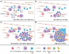
          Chronically inflamed tissues commonly accrue lymphocyte aggregates that facilitate local T cell-B cell interactions. These aggregates can range from small, loosely arranged lymphocyte clusters to large, organized ectopic lymphoid structures. In some cases, ectopic lymphoid structures develop germinal centers that house prototypical T follicular helper (Tfh) cells with high expression of Bcl6, CXCR5, PD-1, and ICOS. However, in many chronically inflamed tissues, the T cells that interact with B cells show substantial differences from Tfh cells in their surface phenotypes, migratory capacity, and transcriptional regulation. This review discusses observations from multiple diseases and models in which tissue-infiltrating T cells produce factors associated with B cell help, including IL-21 and the B cell chemoattractant CXCL13, yet vary dramatically in their resemblance to Tfh cells. Particular attention is given to the PD-1 hi CXCR5 Bcl6 low T peripheral helper (Tph) cell population in rheumatoid arthritis, which infiltrates inflamed synovium through expression of chemokine receptors such as CCR2 and augments synovial B cell responses via CXCL13 and IL-21. The factors that regulate CD4 + T cell production of CXCL13 and IL-21 in these settings are also discussed. Understanding the range of T cell populations that can provide help to B cells within chronically inflamed tissues is essential to recognize these cells in diverse inflammatory conditions and to optimize either broad or selective therapeutic targeting of B cell-helper T cells.

          Related collections

          Most cited references86

          • Record: found
          • Abstract: found
          • Article: not found

          Meningeal B-cell follicles in secondary progressive multiple sclerosis associate with early onset of disease and severe cortical pathology.

          Intrathecal antibody production is a hallmark of multiple sclerosis and humoral immunity is thought to play an important role in the inflammatory response and development of demyelinated lesions. The presence of lymphoid follicle-like structures in the cerebral meninges of some multiple sclerosis patients indicates that B-cell maturation can be sustained locally within the CNS and contribute to the establishment of a compartmentalized humoral immune response. In this study we examined the distribution of ectopic B-cell follicles in multiple sclerosis cases with primary and secondary progressive clinical courses to determine their association with clinical and neuropathological features. A detailed immunohistochemical and morphometric analysis was performed on post-mortem brain tissue samples from 29 secondary progressive (SP) and 7 primary progressive (PP) multiple sclerosis cases. B-cell follicles were detected in the meninges entering the cerebral sulci of 41.4% of the SPMS cases, but not in PPMS cases. The SPMS cases with follicles significantly differed from those without with respect to a younger age at multiple sclerosis onset, irreversible disability and death and more pronounced demyelination, microglia activation and loss of neurites in the cerebral cortex. Cortical demyelination in these SPMS cases was also more severe than in PPMS cases. Notably, all meningeal B-cell follicles were found adjacent to large subpial cortical lesions, suggesting that soluble factors diffusing from these structures have a pathogenic role. These data support an immunopathogenetic mechanism whereby B-cell follicles developing in the multiple sclerosis meninges exacerbate the detrimental effects of humoral immunity with a subsequent major impact on the integrity of the cortical structures.
            Bookmark
            • Record: found
            • Abstract: found
            • Article: not found

            Follicular B Helper T Cells Express Cxc Chemokine Receptor 5, Localize to B Cell Follicles, and Support Immunoglobulin Production

            Chemokines and their receptors have been identified as major regulators controlling the functional organization of secondary lymphoid organs. Here we show that expression of CXC chemokine receptor 5 (CXCR5), a chemokine receptor required for B cell homing to B cell follicles, defines a novel subpopulation of B helper T cells localizing to follicles. In peripheral blood these cells coexpress CD45RO and the T cell homing CC chemokine receptor 7 (CCR7). In secondary lymphoid organs, CD4+CXCR5+ cells lose expression of CCR7, which allows them to localize to B cell follicles and germinal centers where they express high levels of CD40 ligand (CD40L), a costimulatory molecule required for B cell activation and inducible costimulator (ICOS), a recently identified costimulatory molecule of the CD28 family. Thus, when compared with CD4+CD45RO+CXCR5− cells, CD4+CD45RO+CXCR5+ tonsillar T cells efficiently support the production of immunoglobulin (Ig)A and IgG. In contrast, analysis of the memory response revealed that long-lasting memory cells are found within the CD4+CD45RO+CXCR5− population, suggesting that CXCR5+CD4 cells represent recently activated effector cells. Based on the characteristic localization within secondary lymphoid organs, we suggest to term these cells “follicular B helper T cells” (TFH).
              Bookmark
              • Record: found
              • Abstract: found
              • Article: not found

              Cxc Chemokine Receptor 5 Expression Defines Follicular Homing T Cells with B Cell Helper Function

              Leukocyte traffic through secondary lymphoid tissues is finely tuned by chemokines. We have studied the functional properties of a human T cell subset marked by the expression of CXC chemokine receptor 5 (CXCR5). Memory but not naive T cells from tonsils are CXCR5+ and migrate in response to the B cell–attracting chemokine 1 (BCA-1), which is selectively expressed by reticular cells and blood vessels within B cell follicles. Tonsillar CXCR5+ T cells do not respond to other chemokines present in secondary lymphoid tissues, including secondary lymphoid tissue chemokine (SLC), EBV-induced molecule 1 ligand chemokine (ELC), and stromal cell–derived factor 1 (SDF-1). The involvement of tonsillar CXCR5+ T cells in humoral immune responses is suggested by their localization in the mantle and light zone germinal centers of B cell follicles and by the concomitant expression of activation and costimulatory markers, including CD69, HLA-DR, and inducible costimulator (ICOS). Peripheral blood CXCR5+ T cells also belong to the CD4+ memory T cell subset but, in contrast to tonsillar cells, are in a resting state and migrate weakly to chemokines. CXCR5+ T cells are very inefficient in the production of cytokines but potently induce antibody production during coculture with B cells. These properties portray CXCR5+ T cells as a distinct memory T cell subset with B cell helper function, designated here as follicular B helper T cells (TFH).
                Bookmark

                Author and article information

                Contributors
                Journal
                Front Immunol
                Front Immunol
                Front. Immunol.
                Frontiers in Immunology
                Frontiers Media S.A.
                1664-3224
                23 August 2018
                2018
                : 9
                : 1924
                Affiliations
                Division of Rheumatology, Immunology, Allergy, Department of Medicine, Brigham and Women's Hospital, Harvard Medical School , Boston, MA, United States
                Author notes

                Edited by: Maria Pia Cicalese, San Raffaele Scientific Institute (IRCCS), Italy

                Reviewed by: Constantinos Petrovas, National Institutes of Health (NIH), United States; Thomas Ciucci, National Cancer Institute (NCI), United States

                *Correspondence: Deepak A. Rao darao@ 123456bwh.harvard.edu

                This article was submitted to T Cell Biology, a section of the journal Frontiers in Immunology

                Article
                10.3389/fimmu.2018.01924
                6115497
                30190721
                4a1c8d2e-5926-49b3-8be7-16b295262d1f
                Copyright © 2018 Rao.

                This is an open-access article distributed under the terms of the Creative Commons Attribution License (CC BY). The use, distribution or reproduction in other forums is permitted, provided the original author(s) and the copyright owner(s) are credited and that the original publication in this journal is cited, in accordance with accepted academic practice. No use, distribution or reproduction is permitted which does not comply with these terms.

                History
                : 30 May 2018
                : 06 August 2018
                Page count
                Figures: 2, Tables: 0, Equations: 0, References: 127, Pages: 13, Words: 11739
                Funding
                Funded by: National Institute of Arthritis and Musculoskeletal and Skin Diseases 10.13039/100000069
                Award ID: K08AR072791-01
                Funded by: Rheumatology Research Foundation 10.13039/100006260
                Funded by: Brigham and Women's Hospital 10.13039/100005292
                Categories
                Immunology
                Review

                Immunology
                t follicular helper cells,t peripheral helper cells,il-21,cxcl13,b cells,ectopic lymphoid structure,ectopic lymphoid follicle,tertiary lymphoid tissue

                Comments

                Comment on this article