2
views
0
recommends
+1 Recommend
0 collections
    0
    shares
      • Record: found
      • Abstract: found
      • Article: found
      Is Open Access

      Lipophagy deficiency exacerbates ectopic lipid accumulation and tubular cells injury in diabetic nephropathy

      research-article

      Read this article at

      Bookmark
          There is no author summary for this article yet. Authors can add summaries to their articles on ScienceOpen to make them more accessible to a non-specialist audience.

          Abstract

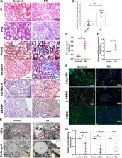
          Autophagy-mediated lipotoxicity plays a critical role in the progression of diabetic nephropathy (DN), but the precise mechanism is not fully understood. Whether lipophagy, a selective type of autophagy participates in renal ectopic lipid deposition (ELD) and lipotoxicity in the kidney of DN is unknown. Here, decreased lipophagy, increased ELD and lipotoxcity were observed in tubular cells of patients with DN, which were accompanied with reduced expression of AdipoR1 and p-AMPK. Similar results were found in db/db mice, these changes were reversed by AdipoRon, an adiponectin receptor activator that promotes autophagy. Additionally, a significantly decreased level of lipophagy was observed in HK-2 cells, a human proximal tubular cell line treated with high glucose, which was consistent with increased lipid deposition, apoptosis and fibrosis, while were partially alleviated by AdipoRon. However, these effects were abolished by pretreatment with ULK1 inhibitor SBI-0206965, autophagy inhibitor chloroquine and enhanced by AMPK activator AICAR. These data suggested by the first time that autophagy-mediated lipophagy deficiency plays a critical role in the ELD and lipid-related renal injury of DN.

          Related collections

          Most cited references48

          • Record: found
          • Abstract: found
          • Article: not found

          AMPK and mTOR regulate autophagy through direct phosphorylation of Ulk1.

          Autophagy is a process by which components of the cell are degraded to maintain essential activity and viability in response to nutrient limitation. Extensive genetic studies have shown that the yeast ATG1 kinase has an essential role in autophagy induction. Furthermore, autophagy is promoted by AMP activated protein kinase (AMPK), which is a key energy sensor and regulates cellular metabolism to maintain energy homeostasis. Conversely, autophagy is inhibited by the mammalian target of rapamycin (mTOR), a central cell-growth regulator that integrates growth factor and nutrient signals. Here we demonstrate a molecular mechanism for regulation of the mammalian autophagy-initiating kinase Ulk1, a homologue of yeast ATG1. Under glucose starvation, AMPK promotes autophagy by directly activating Ulk1 through phosphorylation of Ser 317 and Ser 777. Under nutrient sufficiency, high mTOR activity prevents Ulk1 activation by phosphorylating Ulk1 Ser 757 and disrupting the interaction between Ulk1 and AMPK. This coordinated phosphorylation is important for Ulk1 in autophagy induction. Our study has revealed a signalling mechanism for Ulk1 regulation and autophagy induction in response to nutrient signalling.
            Bookmark
            • Record: found
            • Abstract: found
            • Article: not found

            Autophagy regulates lipid metabolism.

            The intracellular storage and utilization of lipids are critical to maintain cellular energy homeostasis. During nutrient deprivation, cellular lipids stored as triglycerides in lipid droplets are hydrolysed into fatty acids for energy. A second cellular response to starvation is the induction of autophagy, which delivers intracellular proteins and organelles sequestered in double-membrane vesicles (autophagosomes) to lysosomes for degradation and use as an energy source. Lipolysis and autophagy share similarities in regulation and function but are not known to be interrelated. Here we show a previously unknown function for autophagy in regulating intracellular lipid stores (macrolipophagy). Lipid droplets and autophagic components associated during nutrient deprivation, and inhibition of autophagy in cultured hepatocytes and mouse liver increased triglyceride storage in lipid droplets. This study identifies a critical function for autophagy in lipid metabolism that could have important implications for human diseases with lipid over-accumulation such as those that comprise the metabolic syndrome.
              Bookmark
              • Record: found
              • Abstract: found
              • Article: not found

              Pathologic classification of diabetic nephropathy.

              Although pathologic classifications exist for several renal diseases, including IgA nephropathy, focal segmental glomerulosclerosis, and lupus nephritis, a uniform classification for diabetic nephropathy is lacking. Our aim, commissioned by the Research Committee of the Renal Pathology Society, was to develop a consensus classification combining type1 and type 2 diabetic nephropathies. Such a classification should discriminate lesions by various degrees of severity that would be easy to use internationally in clinical practice. We divide diabetic nephropathy into four hierarchical glomerular lesions with a separate evaluation for degrees of interstitial and vascular involvement. Biopsies diagnosed as diabetic nephropathy are classified as follows: Class I, glomerular basement membrane thickening: isolated glomerular basement membrane thickening and only mild, nonspecific changes by light microscopy that do not meet the criteria of classes II through IV. Class II, mesangial expansion, mild (IIa) or severe (IIb): glomeruli classified as mild or severe mesangial expansion but without nodular sclerosis (Kimmelstiel-Wilson lesions) or global glomerulosclerosis in more than 50% of glomeruli. Class III, nodular sclerosis (Kimmelstiel-Wilson lesions): at least one glomerulus with nodular increase in mesangial matrix (Kimmelstiel-Wilson) without changes described in class IV. Class IV, advanced diabetic glomerulosclerosis: more than 50% global glomerulosclerosis with other clinical or pathologic evidence that sclerosis is attributable to diabetic nephropathy. A good interobserver reproducibility for the four classes of DN was shown (intraclass correlation coefficient = 0.84) in a test of this classification.
                Bookmark

                Author and article information

                Contributors
                sunlin@csu.edu.cn
                Journal
                Cell Death Dis
                Cell Death Dis
                Cell Death & Disease
                Nature Publishing Group UK (London )
                2041-4889
                30 October 2021
                30 October 2021
                November 2021
                : 12
                : 11
                : 1031
                Affiliations
                [1 ]GRID grid.452708.c, ISNI 0000 0004 1803 0208, Department of Nephrology, , The Second Xiangya Hospital, Central South University, Key Laboratory of Kidney Disease and Blood Purification, ; Changsha, Hunan China
                [2 ]GRID grid.431010.7, Department of Nephrology, , The Third Xiangya Hospital, Central South University, ; Changsha, Hunan China
                Author information
                http://orcid.org/0000-0002-6552-0176
                http://orcid.org/0000-0002-4544-0822
                Article
                4326
                10.1038/s41419-021-04326-y
                8557213
                34718329
                4a441a0c-525f-4860-91bb-76118ba188d7
                © The Author(s) 2021

                Open Access This article is licensed under a Creative Commons Attribution 4.0 International License, which permits use, sharing, adaptation, distribution and reproduction in any medium or format, as long as you give appropriate credit to the original author(s) and the source, provide a link to the Creative Commons license, and indicate if changes were made. The images or other third party material in this article are included in the article’s Creative Commons license, unless indicated otherwise in a credit line to the material. If material is not included in the article’s Creative Commons license and your intended use is not permitted by statutory regulation or exceeds the permitted use, you will need to obtain permission directly from the copyright holder. To view a copy of this license, visit http://creativecommons.org/licenses/by/4.0/.

                History
                : 20 April 2021
                : 14 October 2021
                : 15 October 2021
                Funding
                Funded by: FundRef https://doi.org/10.13039/501100011002, National Science Foundation of China | National Natural Science Foundation of China-Yunnan Joint Fund (NSFC-Yunnan Joint Fund);
                Award ID: 81730018
                Award Recipient :
                Funded by: the National Key R&D Program of China (2016YFC1305501; 2018YFC1314002)
                Categories
                Article
                Custom metadata
                © The Author(s) 2021

                Cell biology
                chronic kidney disease,diabetes complications
                Cell biology
                chronic kidney disease, diabetes complications

                Comments

                Comment on this article