10
views
0
recommends
+1 Recommend
0 collections
    0
    shares
      • Record: found
      • Abstract: found
      • Article: found
      Is Open Access

      IL-38 has an anti-inflammatory action in psoriasis and its expression correlates with disease severity and therapeutic response to anti-IL-17A treatment

      research-article

      Read this article at

      Bookmark
          There is no author summary for this article yet. Authors can add summaries to their articles on ScienceOpen to make them more accessible to a non-specialist audience.

          Abstract

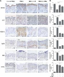
          IL-36 cytokines, a subgroup of IL-1 family, comprise IL-36α, IL-36β, and IL-36γ agonists, abundantly expressed in psoriatic skin, and IL-36RA and IL-38 antagonists. In psoriatic skin, IL-36 cytokines interfere with keratinocyte cornification programs and induce the release of antimicrobial peptides and chemokines active on neutrophils and Th17 lymphocytes. To date, the role of IL-38 antagonist in psoriasis remains to be defined. Here, we demonstrate that skin and circulating IL-38 levels are reduced in psoriatic patients and in other skin diseases characterized by neutrophilic infiltrate. In psoriasis, the balance of IL-36γ agonist/IL-38 antagonist serum levels is in favor of agonists and is closely associated with disease severity. Interestingly, IL-38 is upregulated by anti-IL-17A biological treatment and positively correlates with the therapeutic efficacy of secukinumab in psoriatic patients. The downregulation of IL-38 expression is strictly related to keratinocyte de-differentiation triggered by the inflammatory cytokines IL-36γ, IL-17, and IL-22. Finally, we demonstrate that administration of recombinant full-length IL-38 counteracts in vitro the biological processes induced by IL-36γ in human keratinocytes and endothelial cells and attenuates in vivo the severity of the psoriasiform phenotype induced by IMQ in mice. Such effects are achieved by restoring the physiological programs of keratinocyte proliferation and differentiation, and reducing the immune cell infiltrates.

          Related collections

          Most cited references42

          • Record: found
          • Abstract: found
          • Article: not found

          Interleukin-36-receptor antagonist deficiency and generalized pustular psoriasis.

          Generalized pustular psoriasis is a life-threatening disease of unknown cause. It is characterized by sudden, repeated episodes of high-grade fever, generalized rash, and disseminated pustules, with hyperleukocytosis and elevated serum levels of C-reactive protein, which may be associated with plaque-type psoriasis. We performed homozygosity mapping and direct sequencing in nine Tunisian multiplex families with autosomal recessive generalized pustular psoriasis. We assessed the effect of mutations on protein expression and conformation, stability, and function. We identified significant linkage to an interval of 1.2 megabases on chromosome 2q13-q14.1 and a homozygous missense mutation in IL36RN, encoding an interleukin-36-receptor antagonist (interleukin-36Ra), an antiinflammatory cytokine. This mutation predicts the substitution of a proline residue for leucine at amino acid position 27 (L27P). Homology-based structural modeling of human interleukin-36Ra suggests that the proline at position 27 affects both the stability of interleukin-36Ra and its interaction with its receptor, interleukin-1 receptor-like 2 (interleukin-1 receptor-related protein 2). Biochemical analyses showed that the L27P variant was poorly expressed and less potent than the nonvariant interleukin-36Ra in inhibiting a cytokine-induced response in an interleukin-8 reporter assay, leading to enhanced production of inflammatory cytokines (interleukin-8 in particular) by keratinocytes from the patients. Aberrant interleukin-36Ra structure and function lead to unregulated secretion of inflammatory cytokines and generalized pustular psoriasis. (Funded by Agence Nationale de la Recherche and Société Française de Dermatologie.).
            Bookmark
            • Record: found
            • Abstract: found
            • Article: not found

            Mutations in IL36RN/IL1F5 are associated with the severe episodic inflammatory skin disease known as generalized pustular psoriasis.

            Generalized pustular psoriasis (GPP) is a rare and yet potentially lethal clinical variant of psoriasis, characterized by the formation of sterile cutaneous pustules, neutrophilia, fever and features of systemic inflammation. We sequenced the exomes of five unrelated individuals diagnosed with GPP. Nonsynonymous, splice-site, insertion, and deletion variants with an estimated population frequency of T (p.Ser113Leu) missense substitution of IL36RN was identified in two individuals, with a third subject found to be a compound heterozygote for c.338C>T (p.Ser113Leu) and a c.142C>T (p.Arg48Trp) missense substitution. IL36RN (previously known as IL1F5) encodes an IL-1 family receptor antagonist, which opposes the activity of the IL-36A and IL-36G innate cytokines. Homology searches revealed that GPP mutations alter evolutionarily conserved residues. Homozygosity for the c.338C>T (p.Ser113Leu) variant is associated with an elevated proinflammatory response following ex vivo stimulation with IL36A. These findings suggest loss of function of IL36RN as the genetic basis of GPP and implicate innate immune dysregulation in this severe episodic inflammatory disease, thereby highlighting IL-1 signaling as a potential target for therapeutic intervention. Copyright © 2011 The American Society of Human Genetics. Published by Elsevier Inc. All rights reserved.
              Bookmark
              • Record: found
              • Abstract: found
              • Article: not found

              Pathogenesis of psoriasis and development of treatment.

              The pathogenesis of psoriasis can be explained by dysregulation of immunological cell function as well as keratinocyte proliferation/differentiation. Recently, the immunological pathomechanism has been clarified substantially. Whereas T-helper (Th)1 overactivation was thought to induce occurrence of psoriasis, it has been demonstrated that Th17 cells play a key role. Th17 development is maintained by interleukin (IL)-23 mainly produced by dendritic cells. Th17 cells produce various cytokines, including IL-17A, IL-17F and IL-22. IL-17A and IL-22 induce not only keratinocyte proliferation, but also tumor necrosis factor (TNF)-α, chemokine (C-X-C motif) ligand (CXCL)1 and CXCL8 production. TNF-α accelerates the infiltration of inflammatory cells, including lymphocytes, monocytes and neutrophils, from the peripheral blood into skin with dendritic cell activation. In addition, antimicrobial peptides are overexpressed in psoriatic skin lesions, and the antimicrobial peptide, LL-37, activates dendritic cells, which leads to the development of inflammation. Furthermore, activation of nuclear factor-κB signal induces the expression of keratins 6 and 16 in keratinocytes, which are associated with acanthosis and reduced turnover time in the epidermis. The progression of the pathomechanism contributes to the development of new therapies for psoriasis.
                Bookmark

                Author and article information

                Contributors
                +39-06-66464776 , s.madonna@idi.it
                Journal
                Cell Death Dis
                Cell Death Dis
                Cell Death & Disease
                Nature Publishing Group UK (London )
                2041-4889
                30 October 2018
                30 October 2018
                November 2018
                : 9
                : 11
                : 1104
                Affiliations
                [1 ]ISNI 0000 0004 1758 0179, GRID grid.419457.a, Laboratory of Experimental Immunology and Integrated Research Center for PSOriasis (CRI-PSO), , Istituto Dermopatico dell‘Immacolata IDI-IRCCS, ; via Monti di Creta, 104, ROME, Italy
                [2 ]ISNI 0000 0004 1763 1124, GRID grid.5611.3, Section of Dermatology, Department of Medicine, , University of Verona, ; P.zza Stefani, 1, Verona, 37126 Italy
                [3 ]ISNI 0000 0001 0703 675X, GRID grid.430503.1, Department of Biochemistry & Molecular Genetics, School of Medicine, , University of Colorado Denver, Anschutz Campus, ; Aurora, 80045 CO USA
                [4 ]ISNI 0000 0004 1790 0507, GRID grid.429699.9, Istituto di Biostrutture e Bioimmagini-CNR and CIRPEB, ; Via Mezzocannone, 16, Naples, 80134 Italy
                [5 ]ISNI 0000 0004 1758 0179, GRID grid.419457.a, 1st Division of Dermatology and CRI-PSO, , Istituto Dermopatico dell‘Immacolata IDI-IRCCS, ; via Monti di Creta, 104, Rome, 00167 Italy
                [6 ]ISNI 0000 0004 1758 0179, GRID grid.419457.a, CRI-PSO Istituto Dermopatico dell’Immacolata, , IDI-IRCCS, ; via Monti di Creta, 104, Rome, 00167 Italy
                [7 ]INMP/NIHMP, via di S.Gallicano, 25, Rome, 00153 Italy
                [8 ]ISNI 0000 0004 0444 9382, GRID grid.10417.33, Department of Medicine, , Radboud University Medical Center, ; 6525 HP Nijmegen, The Netherlands
                [9 ]ISNI 0000 0001 0703 675X, GRID grid.430503.1, Department of Medicine, School of Medicine, , University of Colorado, Denver, Anschutz Campus, ; Aurora, CO USA
                Author information
                http://orcid.org/0000-0001-5997-756X
                http://orcid.org/0000-0002-7537-6833
                Article
                1143
                10.1038/s41419-018-1143-3
                6207563
                30377293
                4b7d1c0d-9dbc-43e9-9cf5-40256a7c53fc
                © The Author(s) 2018

                Open Access This article is licensed under a Creative Commons Attribution 4.0 International License, which permits use, sharing, adaptation, distribution and reproduction in any medium or format, as long as you give appropriate credit to the original author(s) and the source, provide a link to the Creative Commons license, and indicate if changes were made. The images or other third party material in this article are included in the article’s Creative Commons license, unless indicated otherwise in a credit line to the material. If material is not included in the article’s Creative Commons license and your intended use is not permitted by statutory regulation or exceeds the permitted use, you will need to obtain permission directly from the copyright holder. To view a copy of this license, visit http://creativecommons.org/licenses/by/4.0/.

                History
                : 1 June 2018
                : 10 September 2018
                : 8 October 2018
                Categories
                Article
                Custom metadata
                © The Author(s) 2018

                Cell biology
                Cell biology

                Comments

                Comment on this article