12
views
0
recommends
+1 Recommend
0 collections
    0
    shares
      • Record: found
      • Abstract: found
      • Article: found
      Is Open Access

      Systemic administration of the di-apocarotenoid norbixin (BIO201) is neuroprotective, preserves photoreceptor function and inhibits A2E and lipofuscin accumulation in animal models of age-related macular degeneration and Stargardt disease

      research-article

      Read this article at

      Bookmark
          There is no author summary for this article yet. Authors can add summaries to their articles on ScienceOpen to make them more accessible to a non-specialist audience.

          Abstract

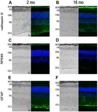
          Atrophic A\age-related macular degeneration (AMD) and Stargardt disease (STGD) are major blinding diseases affecting millions of patients worldwide, but no treatment is available. In dry AMD and STGD oxidative stress and subretinal accumulation of N-retinylidene- N-retinylethanolamine (A2E), a toxic by-product of the visual cycle, causes retinal pigment epithelium (RPE) and photoreceptor degeneration leading to visual impairment. Acute and chronic retinal degeneration following blue light damage (BLD) in BALB/c mice and aging of Abca4 -/- Rdh8 -/- mice, respectively, reproduce features of AMD and STGD. Efficacy of systemic administrations of 9'- cis-norbixin (norbixin), a natural di-apocarotenoid, prepared from Bixa orellana seeds with anti-oxidative properties, was evaluated during BLD in BALB/c mice, and in Abca4 -/- Rdh8 -/- mice of different ages, following three experimental designs: “preventive”, “early curative” and “late curative” supplementations. Norbixin injected intraperitoneally in BALB/c mice, maintained scotopic and photopic electroretinogram amplitude and was neuroprotective. Norbixin chronic oral administration for 6 months in Abca4 -/- Rdh8 -/- mice following the “early curative” supplementation showed optimal neuroprotection and maintenance of photoreceptor function and reduced ocular A2E accumulation. Thus, norbixin appears promising as a systemic drug candidate for both AMD and STGD treatment.

          Related collections

          Most cited references55

          • Record: found
          • Abstract: found
          • Article: not found

          The Rd8 mutation of the Crb1 gene is present in vendor lines of C57BL/6N mice and embryonic stem cells, and confounds ocular induced mutant phenotypes.

          We noted an unexpected inheritance pattern of lesions in several strains of gene-manipulated mice with ocular phenotypes. The lesions, which appeared at various stages of backcross to C57BL/6, bore resemblance to the rd8 retinal degeneration phenotype. We set out to examine the prevalence of this mutation in induced mutant mouse lines, vendor C57BL/6 mice and in widely used embryonic stem cells. Ocular lesions were evaluated by fundus examination and histopathology. Detection of the rd8 mutation at the genetic level was performed by PCR with appropriate primers. Data were confirmed by DNA sequencing in selected cases. Analysis of several induced mutant mouse lines with ocular disease phenotypes revealed that the disease was associated 100% with the presence of the rd8 mutation in the Crb1 gene rather than with the gene of interest. DNA analysis of C57BL/6 mice from common commercial vendors demonstrated the presence of the rd8 mutation in homozygous form in all C57BL/6N substrains, but not in the C57BL/6J substrain. A series of commercially available embryonic stem cells of C57BL/6N origin and C57BL/6N mouse lines used to generate ES cells also contained the rd8 mutation. Affected mice displayed ocular lesions typical of rd8, which were detectable by funduscopy and histopathology as early as 6 weeks of age. These findings identify the presence of the rd8 mutation in the C57BL/6N mouse substrain used widely to produce transgenic and knockout mice. The results have grave implications for the vision research community who develop mouse lines to study eye disease, as presence of rd8 can produce significant disease phenotypes unrelated to the gene or genes of interest. It is suggested that researchers screen for rd8 if their mouse lines were generated on the C57BL/6N background, bear resemblance to the rd8 phenotype, or are of indeterminate origin.
            Bookmark
            • Record: found
            • Abstract: found
            • Article: not found

            Oxidative damage-induced inflammation initiates age-related macular degeneration.

            Oxidative damage and inflammation are postulated to be involved in age-related macular degeneration (AMD). However, the molecular signal(s) linking oxidation to inflammation in this late-onset disease is unknown. Here we describe AMD-like lesions in mice after immunization with mouse serum albumin adducted with carboxyethylpyrrole, a unique oxidation fragment of docosahexaenoic acid that has previously been found adducting proteins in drusen from AMD donor eye tissues and in plasma samples from individuals with AMD. Immunized mice develop antibodies to this hapten, fix complement component-3 in Bruch's membrane, accumulate drusen below the retinal pigment epithelium during aging, and develop lesions in the retinal pigment epithelium mimicking geographic atrophy, the blinding end-stage condition characteristic of the dry form of AMD. We hypothesize that these mice are sensitized to the generation of carboxyethylpyrrole adducts in the outer retina, where docosahexaenoic acid is abundant and conditions for oxidative damage are permissive. This new model provides a platform for dissecting the molecular pathology of oxidative damage in the outer retina and the immune response contributing to AMD.
              Bookmark
              • Record: found
              • Abstract: found
              • Article: not found

              Photoreceptor loss in age-related macular degeneration.

              The authors showed previously that parafoveal rods, but not cones, decrease during the course of adulthood in donor eyes that were screened to exclude the grossly visible macular drusen and pigmentary disturbances typical of age-related macular degeneration (AMD). Because AMD begins in the parafovea, this selective loss of rods actually may be subclinical AMD not yet visible in the fundus. If so, AMD must have a predilection for rods over cones. The authors tested this hypothesis by determining the relative numbers of cones and rods in donor eyes with mid-to late-stage AMD and in age-matched controls. Thirteen eyes (from seven donors) with grossly visible macular drusen and pigmentary disturbances were either wholemounted for photoreceptor counts or sectioned through the fovea for histopathology and carbonic anhydrase histochemistry to label red-green cones. Eyes were assigned to AMD or control groups on the basis of histopathology and clinical history. Five nonexudative AMD (NE-AMD) eyes from three donors showed sparing of foveal cones and loss of rods and cones in the parafovea. In two donors, rod loss exceeded cone loss at most parafoveal locations, and in one donor, rod density was normal and cone density was reduced. In eight exudative AMD (EX-AMD) eyes from five donors, photoreceptors surviving along the margins of and overlying disciform scars were largely cones. Photoreceptors are lost in NE-AMD as well as in the more severe exudative form, consistent with functional and clinical studies. The authors propose that rods die in older eyes without evidence of overt retinal pigment epithelial disease. In persons susceptible to AMD, the retinal pigment epithelium becomes dysfunctional. Secondarily, rod loss continues and cones begin to degenerate. Eventually, only degenerate cones remain; ultimately, all photoreceptors may disappear.
                Bookmark

                Author and article information

                Journal
                Aging (Albany NY)
                Aging (Albany NY)
                Aging
                Aging (Albany NY)
                Impact Journals
                1945-4589
                15 April 2020
                07 April 2020
                : 12
                : 7
                : 6151-6171
                Affiliations
                [1 ]Sorbonne Université, INSERM, CNRS, Institut de la Vision, Paris 75012, France
                [2 ]Biophytis, Sorbonne Université, Campus Pierre and Marie Curie, Paris 75005, France
                Author notes
                Correspondence to: Serge Camelo; email: serge.camelo@biophytis.com
                Article
                103014 103014
                10.18632/aging.103014
                7185133
                32255762
                4ca22542-49cf-4ab4-9a45-79f359ca4b64
                Copyright © 2020 Fontaine et al.

                This is an open-access article distributed under the terms of the Creative Commons Attribution License (CC BY 3.0), which permits unrestricted use, distribution, and reproduction in any medium, provided the original author and source are credited.

                History
                : 05 November 2019
                : 02 March 2020
                Categories
                Research Paper

                Cell biology
                norbixin,retinal function,a2e,amd,stargardt disease
                Cell biology
                norbixin, retinal function, a2e, amd, stargardt disease

                Comments

                Comment on this article