3
views
0
recommends
+1 Recommend
0 collections
    0
    shares
      • Record: found
      • Abstract: found
      • Article: found
      Is Open Access

      The Clinical Significance of Iron Overload and Iron Metabolism in Myelodysplastic Syndrome and Acute Myeloid Leukemia

      review-article

      Read this article at

      Bookmark
          There is no author summary for this article yet. Authors can add summaries to their articles on ScienceOpen to make them more accessible to a non-specialist audience.

          Abstract

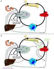
          Myelodysplasticsyndrome (MDS) and acute myeloid leukemia (AML) are clonal hematopoietic stem cell diseases leading to an insufficient formation of functional blood cells. Disease-immanent factors as insufficient erythropoiesis and treatment-related factors as recurrent treatment with red blood cell transfusions frequently lead to systemic iron overload in MDS and AML patients. In addition, alterations of function and expression of proteins associated with iron metabolism are increasingly recognized to be pathogenetic factors and potential vulnerabilities of these diseases. Iron is known to be involved in multiple intracellular and extracellular processes. It is essential for cell metabolism as well as for cell proliferation and closely linked to the formation of reactive oxygen species. Therefore, iron can influence the course of clonal myeloid disorders, the leukemic environment and the occurrence as well as the defense of infections. Imbalances of iron homeostasis may induce cell death of normal but also of malignant cells. New potential treatment strategies utilizing the importance of the iron homeostasis include iron chelation, modulation of proteins involved in iron metabolism, induction of leukemic cell death via ferroptosis and exploitation of iron proteins for the delivery of antileukemic drugs. Here, we provide an overview of some of the latest findings about the function, the prognostic impact and potential treatment strategies of iron in patients with MDS and AML.

          Related collections

          Most cited references198

          • Record: found
          • Abstract: found
          • Article: not found

          Ferroptosis: an iron-dependent form of nonapoptotic cell death.

          Nonapoptotic forms of cell death may facilitate the selective elimination of some tumor cells or be activated in specific pathological states. The oncogenic RAS-selective lethal small molecule erastin triggers a unique iron-dependent form of nonapoptotic cell death that we term ferroptosis. Ferroptosis is dependent upon intracellular iron, but not other metals, and is morphologically, biochemically, and genetically distinct from apoptosis, necrosis, and autophagy. We identify the small molecule ferrostatin-1 as a potent inhibitor of ferroptosis in cancer cells and glutamate-induced cell death in organotypic rat brain slices, suggesting similarities between these two processes. Indeed, erastin, like glutamate, inhibits cystine uptake by the cystine/glutamate antiporter (system x(c)(-)), creating a void in the antioxidant defenses of the cell and ultimately leading to iron-dependent, oxidative death. Thus, activation of ferroptosis results in the nonapoptotic destruction of certain cancer cells, whereas inhibition of this process may protect organisms from neurodegeneration. Copyright © 2012 Elsevier Inc. All rights reserved.
            Bookmark
            • Record: found
            • Abstract: found
            • Article: not found

            Ferroptosis: A Regulated Cell Death Nexus Linking Metabolism, Redox Biology, and Disease

            Ferroptosis is a form of regulated cell death characterized by the iron-dependent accumulation of lipid hydroperoxides to lethal levels. Emerging evidence suggests that ferroptosis represents an ancient vulnerability caused by the incorporation of polyunsaturated fatty acids into cellular membranes, and cells have developed complex systems that exploit and defend against this vulnerability in different contexts. The sensitivity to ferroptosis is tightly linked to numerous biological processes, including amino acid, iron, and polyunsaturated fatty acid metabolism, and the biosynthesis of glutathione, phospholipids, NADPH, and coenzyme Q10. Ferroptosis has been implicated in the pathological cell death associated with degenerative diseases (i.e., Alzheimer's, Huntington's, and Parkinson's diseases), carcinogenesis, stroke, intracerebral hemorrhage, traumatic brain injury, ischemia-reperfusion injury, and kidney degeneration in mammals and is also implicated in heat stress in plants. Ferroptosis may also have a tumor-suppressor function that could be harnessed for cancer therapy. This Primer reviews the mechanisms underlying ferroptosis, highlights connections to other areas of biology and medicine, and recommends tools and guidelines for studying this emerging form of regulated cell death.
              Bookmark
              • Record: found
              • Abstract: found
              • Article: found
              Is Open Access

              Pharmacological inhibition of cystine–glutamate exchange induces endoplasmic reticulum stress and ferroptosis

              Exchange of extracellular cystine for intracellular glutamate by the antiporter system xc − is implicated in numerous pathologies. Pharmacological agents that inhibit system xc − activity with high potency have long been sought, but have remained elusive. In this study, we report that the small molecule erastin is a potent, selective inhibitor of system xc −. RNA sequencing revealed that inhibition of cystine–glutamate exchange leads to activation of an ER stress response and upregulation of CHAC1, providing a pharmacodynamic marker for system xc − inhibition. We also found that the clinically approved anti-cancer drug sorafenib, but not other kinase inhibitors, inhibits system xc − function and can trigger ER stress and ferroptosis. In an analysis of hospital records and adverse event reports, we found that patients treated with sorafenib exhibited unique metabolic and phenotypic alterations compared to patients treated with other kinase-inhibiting drugs. Finally, using a genetic approach, we identified new genes dramatically upregulated in cells resistant to ferroptosis. DOI: http://dx.doi.org/10.7554/eLife.02523.001
                Bookmark

                Author and article information

                Contributors
                Journal
                Front Immunol
                Front Immunol
                Front. Immunol.
                Frontiers in Immunology
                Frontiers Media S.A.
                1664-3224
                19 February 2021
                2020
                : 11
                : 627662
                Affiliations
                [1] 1 Department of Medicine, Hematology/Oncology, University Hospital Frankfurt, Goethe University , Frankfurt am Main, Germany
                [2] 2 German Cancer Consortium (DKTK), Partner Site Frankfurt/Mainz and German Cancer Research Center (DKFZ) , Heidelberg, Germany
                [3] 3 Frankfurt Cancer Institute, Goethe University , Frankfurt am Main, Germany
                Author notes

                Edited by: Stefania Recalcati, University of Milan, Italy

                Reviewed by: Carlo Finelli, University Hospital of Bologna Policlinico S. Orsola-Malpighi, Italy; Norbert Gattermann, Heinrich Heine University of Düsseldorf, Germany

                *Correspondence: Sarah Weber, sarah.weber@ 123456kgu.de ; Hubert Serve, serve@ 123456em.uni-frankfurt.de

                †These authors have contributed equally to this work

                This article was submitted to Molecular Innate Immunity, a section of the journal Frontiers in Immunology

                Article
                10.3389/fimmu.2020.627662
                7933218
                33679722
                4e0885e7-b5e8-4fe3-8ef3-9791f1688b74
                Copyright © 2021 Weber, Parmon, Kurrle, Schnütgen and Serve

                This is an open-access article distributed under the terms of the Creative Commons Attribution License (CC BY). The use, distribution or reproduction in other forums is permitted, provided the original author(s) and the copyright owner(s) are credited and that the original publication in this journal is cited, in accordance with accepted academic practice. No use, distribution or reproduction is permitted which does not comply with these terms.

                History
                : 09 November 2020
                : 31 December 2020
                Page count
                Figures: 3, Tables: 0, Equations: 0, References: 199, Pages: 18, Words: 9625
                Categories
                Immunology
                Review

                Immunology
                myelodysplastic syndrome,acute myeloid leukemia,iron overload,reactive oxygen species,microenvironment,iron chelation

                Comments

                Comment on this article