16
views
0
recommends
+1 Recommend
0 collections
    0
    shares
      • Record: found
      • Abstract: found
      • Article: found
      Is Open Access

      Male versus female skin: What dermatologists and cosmeticians should know

      review-article
      , MD, MPH a , , MD, PhD b , , MD a , , MD, PhD b , , MD a , *
      International Journal of Women's Dermatology
      Elsevier

      Read this article at

      Bookmark
          There is no author summary for this article yet. Authors can add summaries to their articles on ScienceOpen to make them more accessible to a non-specialist audience.

          Abstract

          Introduction

          The skin is important for the perception of health and beauty. Knowledge of the physiological, chemical, and biophysical differences between the skin of male and female patients helps dermatologists develop a proper approach not only for the management of skin diseases but also to properly take care of cosmetic issues. The influence of genetic and environmental factors on skin characteristics is also critical to consider.

          Methods

          A literature search of PubMed and Google was conducted to compare the biophysical and biomechanical properties of the skin of male and female patients using the keywords "skin", "hydration", "water loss", "sebum", "circulation", "color", "thickness", "elasticity", "pH", "friction", "wrinkle", "sex", "male", and "female".

          Results

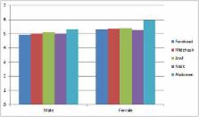
          A total of 1070 titles were found. After removing duplications and non-English papers, the number was reduced to 632. Of the 632 titles, 57 were deemed suitable for inclusion in this review. The studies show that the skin parameters of hydration, transepidermal water loss, sebum, microcirculation, pigmentation, and thickness are generally higher in men but skin pH is higher in women.

          Conclusions

          These parameters can be considered as age markers in some cases and are susceptible to change according to environment and life style. Biometrological studies of the skin provide useful information in the selection of active principles and other ingredients of formulations to develop a specific approach for cosmetic treatments.

          Related collections

          Most cited references57

          • Record: found
          • Abstract: found
          • Article: not found

          Epidermal thickness at different body sites: relationship to age, gender, pigmentation, blood content, skin type and smoking habits.

          Epidermal thickness and its relationship to age, gender, skin type, pigmentation, blood content, smoking habits and body site is important in dermatologic research and was investigated in this study. Biopsies from three different body sites of 71 human volunteers were obtained, and thickness of the stratum corneum and cellular epidermis was measured microscopically using a preparation technique preventing tissue damage. Multiple regressions analysis was used to evaluate the effect of the various factors independently of each other. Mean (SD) thickness of the stratum corneum was 18.3 (4.9) microm at the dorsal aspect of the forearm, 11.0 (2.2) microm at the shoulder and 14.9 (3.4) microm at the buttock. Corresponding values for the cellular epidermis were 56.6 (11.5) microm, 70.3 (13.6) microm and 81.5 (15.7) microm, respectively. Body site largely explains the variation in epidermal thickness, but also a significant individual variation was observed. Thickness of the stratum corneum correlated positively to pigmentation (p = 0.0008) and negatively to the number of years of smoking (p < 0.0001). Thickness of the cellular epidermis correlated positively to blood content (P = 0.028) and was greater in males than in females (P < 0.0001). Epidermal thickness was not correlated to age or skin type.
            Bookmark
            • Record: found
            • Abstract: found
            • Article: not found

            The influence of age and sex on skin thickness, skin collagen and density.

            Forearm skin collagen, dermal thickness and collagen density were measured in a large number of normal subjects as a standard reference for future studies. Skin collagen decreased with age and was less in the females at all ages. There is a direct relationship between skin collagen and dermal thickness but variations in collagen density in disease limit the use of dermal thickness as a guide to changes in its collagen content.
              Bookmark
              • Record: found
              • Abstract: found
              • Article: not found

              Number of cell layers of the stratum corneum in normal skin - relationship to the anatomical location on the body, age, sex and physical parameters.

              We counted the number of cell layers in the stratum corneum (SC) of normal skin taken from different anatomical locations of the body of 301 individuals of various ages. Frozen 6 microm thick sections were stained with a 1% aqueous solution of safranin and observed under a microscope after application of 2% KOH solution. There were great variations in the number of SC cell layers (mean +/- SD) according to location and among different individuals. The smallest number was found in genital skin (6 +/- 2), followed in order by skin of the face (9 +/- 2), neck (10 +/- 2), scalp (12 +/- 2), trunk (13 +/- 4), extremities (15 +/- 4) and the palms and soles (47 +/- 24). The heel showed the largest number (86 +/- 36). No definite correlation was found between the number of corneocyte layers and sex of the individual, whereas there was a slight increase in the number of SC layers with age in the skin of the cheek and back, particularly in male individuals. Comparison of these data with those from functional assessments of the SC of the skin from various locations of healthy adults showed that transepidermal water loss, an indicator of SC barrier function, reflected the number of corneocyte cell layers. In contrast, high-frequency conductance, an indicator of the hydration state of the outer SC, did not seem to be under the influence simply of the number of SC cell layers.
                Bookmark

                Author and article information

                Contributors
                Journal
                Int J Womens Dermatol
                Int J Womens Dermatol
                International Journal of Women's Dermatology
                Elsevier
                2352-6475
                22 June 2018
                September 2018
                22 June 2018
                : 4
                : 3
                : 122-130
                Affiliations
                [a ]Center for Research and Training in Skin Disease and Leprosy, Tehran University of Medical Sciences, Tehran, Iran
                [b ]Research and Studies Center on the Integument, Department of Dermatology, Clinical Investigation Center, Besançon University Hospital, University of Franche-Comté, Besançon, France
                Author notes
                [* ]Corresponding Author. firozali@ 123456sina.tums.ac.ir
                Article
                S2352-6475(18)30013-3
                10.1016/j.ijwd.2018.03.002
                6116811
                30175213
                4e0e6ac1-8584-47be-a097-6902e8f6b0ed
                © 2018 The Authors

                This is an open access article under the CC BY-NC-ND license (http://creativecommons.org/licenses/by-nc-nd/4.0/).

                History
                : 4 August 2017
                : 23 February 2018
                : 4 March 2018
                Categories
                Article

                Comments

                Comment on this article