16
views
0
recommends
+1 Recommend
0 collections
    0
    shares
      • Record: found
      • Abstract: found
      • Article: found
      Is Open Access

      Research Models for Studying Vascular Calcification

      review-article

      Read this article at

      Bookmark
          There is no author summary for this article yet. Authors can add summaries to their articles on ScienceOpen to make them more accessible to a non-specialist audience.

          Abstract

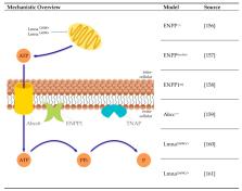
          Calcification of the vessel wall contributes to high cardiovascular morbidity and mortality. Vascular calcification (VC) is a systemic disease with multifaceted contributing and inhibiting factors in an actively regulated process. The exact underlying mechanisms are not fully elucidated and reliable treatment options are lacking. Due to the complex pathophysiology, various research models exist evaluating different aspects of VC. This review aims to give an overview of the cell and animal models used so far to study the molecular processes of VC. Here, in vitro cell culture models of different origins, ex vivo settings using aortic tissue and various in vivo disease-induced animal models are summarized. They reflect different aspects and depict the (patho)physiologic mechanisms within the VC process.

          Related collections

          Most cited references153

          • Record: found
          • Abstract: found
          • Article: not found

          Osteogenic differentiation of purified, culture-expanded human mesenchymal stem cells in vitro.

          Human bone marrow contains a population of cells capable of differentiating along multiple mesenchymal cell lineages. Recently, techniques for the purification and culture-expansion of these human marrow-derived Mesenchymal Stem Cells (MSCs) have been developed. The goals of the current study were to establish a reproducible system for the in vitro osteogenic differentiation of human MSCs, and to characterize the effect of changes in the microenvironment upon the process. MSCs derived from 2nd or 3rd passage were cultured for 16 days in various base media containing 1 to 1000 nM dexamethasone (Dex), 0.01 to 4 mM L-ascorbic acid-2-phosphate (AsAP) or 0.25 mM ascorbic acid, and 1 to 10 mM beta-glycerophosphate (beta GP). Optimal osteogenic differentiation, as determined by osteoblastic morphology, expression of alkaline phosphatase (APase), reactivity with anti-osteogenic cell surface monoclonal antibodies, modulation of osteocalcin mRNA production, and the formation of a mineralized extracellular matrix containing hydroxyapatite was achieved with DMEM base medium plus 100 nM Dex, 0.05 mM AsAP, and 10 mM beta GP. The formation of a continuously interconnected network of APase-positive cells and mineralized matrix supports the characterization of this progenitor population as homogeneous. While higher initial seeding densities did not affect cell number of APase activity, significantly more mineral was deposited in these cultures, suggesting that events which occur early in the differentiation process are linked to end-stage phenotypic expression. Furthermore, cultures allowed to concentrate their soluble products in the media produced more mineralized matrix, thereby implying a role for autocrine or paracrine factors synthesized by human MSCs undergoing osteoblastic lineage progression. This culture system is responsive to subtle manipulations including the basal nutrient medium, dose of physiologic supplements, cell seeding density, and volume of tissue culture medium. Cultured human MSCs provide a useful model for evaluating the multiple factors responsible for the step-wise progression of cells from undifferentiated precursors to secretory osteoblasts, and eventually terminally differentiated osteocytes.
            Bookmark
            • Record: found
            • Abstract: not found
            • Article: not found

            Targeted ablation of Fgf23 demonstrates an essential physiological role of FGF23 in phosphate and vitamin D metabolism

              Bookmark
              • Record: found
              • Abstract: found
              • Article: not found

              Endothelial-to-mesenchymal transition drives atherosclerosis progression.

              The molecular mechanisms responsible for the development and progression of atherosclerotic lesions have not been fully established. Here, we investigated the role played by endothelial-to-mesenchymal transition (EndMT) and its key regulator FGF receptor 1 (FGFR1) in atherosclerosis. In cultured human endothelial cells, both inflammatory cytokines and oscillatory shear stress reduced endothelial FGFR1 expression and activated TGF-β signaling. We further explored the link between disrupted FGF endothelial signaling and progression of atherosclerosis by introducing endothelial-specific deletion of FGF receptor substrate 2 α (Frs2a) in atherosclerotic (Apoe(-/-)) mice. When placed on a high-fat diet, these double-knockout mice developed atherosclerosis at a much earlier time point compared with that their Apoe(-/-) counterparts, eventually demonstrating an 84% increase in total plaque burden. Moreover, these animals exhibited extensive development of EndMT, deposition of fibronectin, and increased neointima formation. Additionally, we conducted a molecular and morphometric examination of left main coronary arteries from 43 patients with various levels of coronary disease to assess the clinical relevance of these findings. The extent of coronary atherosclerosis in this patient set strongly correlated with loss of endothelial FGFR1 expression, activation of endothelial TGF-β signaling, and the extent of EndMT. These data demonstrate a link between loss of protective endothelial FGFR signaling, development of EndMT, and progression of atherosclerosis.
                Bookmark

                Author and article information

                Journal
                Int J Mol Sci
                Int J Mol Sci
                ijms
                International Journal of Molecular Sciences
                MDPI
                1422-0067
                23 March 2020
                March 2020
                : 21
                : 6
                : 2204
                Affiliations
                [1 ]Campus Benjamin Franklin, Department of Nephrology, Charité-Universitätsmedizin Berlin, Corporate Member of Freie Universität Berlin, Humboldt-Universtität zu Berlin, and Berlin Institute of Health, Hindenburgdamm 30, 12203 Berlin, Germany; Jaqueline.Herrmann@ 123456charite.de (J.H.); Milen.Babic@ 123456charite.de (M.B.); Markus.Toelle@ 123456charite.de (M.T.); Markus.vanderGiet@ 123456charite.de (M.v.d.G.)
                [2 ]Department of Chemistry, Biochemistry and Pharmacy, Freie Universität Berlin, Königin-Luise-Straβe 2+4, 14195 Berlin, Germany
                Author notes
                Author information
                https://orcid.org/0000-0001-7388-9611
                Article
                ijms-21-02204
                10.3390/ijms21062204
                7139511
                32210002
                55273702-71b8-4b8a-93f1-e6d6e4df211d
                © 2020 by the authors.

                Licensee MDPI, Basel, Switzerland. This article is an open access article distributed under the terms and conditions of the Creative Commons Attribution (CC BY) license ( http://creativecommons.org/licenses/by/4.0/).

                History
                : 02 March 2020
                : 18 March 2020
                Categories
                Review

                Molecular biology
                vascular calcification,in vitro,ex vivo,in vivo
                Molecular biology
                vascular calcification, in vitro, ex vivo, in vivo

                Comments

                Comment on this article