16
views
0
recommends
+1 Recommend
0 collections
    0
    shares
      • Record: found
      • Abstract: found
      • Article: found
      Is Open Access

      Translation of myelin basic protein mRNA in oligodendrocytes is regulated by integrin activation and hnRNP-K

      research-article

      Read this article at

      Bookmark
          There is no author summary for this article yet. Authors can add summaries to their articles on ScienceOpen to make them more accessible to a non-specialist audience.

          Abstract

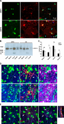
          α6β1-integrin interacts with hnRNP-K, an mRNA-binding protein, during oligodendrocyte differentiation to promote translation of MBP mRNA and myelin synthesis.

          Abstract

          Myelination in the central nervous system provides a unique example of how cells establish asymmetry. The myelinating cell, the oligodendrocyte, extends processes to and wraps multiple axons of different diameter, keeping the number of wraps proportional to the axon diameter. Local regulation of protein synthesis represents one mechanism used to control the different requirements for myelin sheath at each axo–glia interaction. Prior work has established that β1-integrins are involved in the axoglial interactions that initiate myelination. Here, we show that integrin activation regulates translation of a key sheath protein, myelin basic protein (MBP), by reversing the inhibitory effect of the mRNA 3′UTR. During oligodendrocyte differentiation and myelination α6β1-integrin interacts with hnRNP-K, an mRNA-binding protein, which binds to MBP mRNA and translocates from the nucleus to the myelin sheath. Furthermore, knockdown of hnRNP-K inhibits MBP protein synthesis during myelination. Together, these results identify a novel pathway by which axoglial adhesion molecules coordinate MBP synthesis with myelin sheath formation.

          Related collections

          Most cited references40

          • Record: found
          • Abstract: found
          • Article: not found

          Integrin-mediated activation of Cdc42 controls cell polarity in migrating astrocytes through PKCzeta.

          We describe here a signal transduction pathway controlling the establishment of mammalian cell polarity. Scratching a confluent monolayer of primary rat astrocytes leads to polarization of cells at the leading edge. The microtubule organizing center, the microtubule cytoskeleton, and the Golgi reorganize to face the new free space, and directed cell protrusion and migration specifically occur perpendicularly to the scratch. We show here that the interaction of integrins with extracellular matrix at the newly formed cell front leads to the activation and polarized recruitment of Cdc42, which in turn recruits and activates a cytoplasmic mPar6/PKCzeta complex. Localized PKCzeta activity, acting through the microtubule motor protein dynein, is required for all aspects of induced polarity in these cells.
            Bookmark
            • Record: found
            • Abstract: found
            • Article: not found

            Myelin basic protein: a multifunctional protein.

            J Boggs (2006)
            Myelin basic protein (MBP), the second most abundant protein in central nervous system myelin, is responsible for adhesion of the cytosolic surfaces of multilayered compact myelin. A member of the 'intrinsically disordered' or conformationally adaptable protein family, it also appears to have several other functions. It can interact with a number of polyanionic proteins including actin, tubulin, Ca(2+)-calmodulin, and clathrin, and negatively charged lipids, and acquires structure on binding to them. It may act as a membrane actin-binding protein, which might allow it to participate in transmission of extracellular signals to the cytoskeleton in oligodendrocytes and tight junctions in myelin. Some size isoforms of MBP are transported into the nucleus and thus they may also bind polynucleotides. Extracellular signals received by myelin or cultured oligodendrocytes cause changes in phosphorylation of MBP, suggesting that MBP is also involved in signaling. Further study of this very abundant protein will reveal how it is utilized by the oligodendrocyte and myelin for different purposes.
              Bookmark
              • Record: found
              • Abstract: found
              • Article: not found

              Beta1-integrin orients epithelial polarity via Rac1 and laminin.

              Epithelial cells polarize and orient polarity in response to cell-cell and cell-matrix adhesion. Although there has been much recent progress in understanding the general polarizing machinery of epithelia, it is largely unclear how this machinery is controlled by the extracellular environment. To explore the signals from cell-matrix interactions that control orientation of cell polarity, we have used three-dimensional culture systems in which Madin-Darby canine kidney (MDCK) cells form polarized, lumen-containing structures. We show that interaction of collagen I with apical beta1-integrins after collagen overlay of a polarized MDCK monolayer induces activation of Rac1, which is required for collagen overlay-induced tubulocyst formation. Cysts, comprised of a monolayer enclosing a central lumen, form after embedding single cells in collagen. In those cultures, addition of a beta1-integrin function-blocking antibody to the collagen matrix gives rise to cysts that have defects in the organization of laminin into the basement membrane and have inverted polarity. Normal polarity is restored by either expression of activated Rac1, or the inclusion of excess laminin-1 (LN-1). Together, our results suggest a signaling pathway in which the activation of beta1-integrins orients the apical pole of polarized cysts via a mechanism that requires Rac1 activation and laminin organization into the basement membrane.
                Bookmark

                Author and article information

                Journal
                J Cell Biol
                J. Cell Biol
                jcb
                The Journal of Cell Biology
                The Rockefeller University Press
                0021-9525
                1540-8140
                7 March 2011
                : 192
                : 5
                : 797-811
                Affiliations
                [1 ]MRC Centre for Regenerative Medicine and MS Society Translational Research Centre, Centre for Inflammation Research, The Queen’s Medical Research Institute, University of Edinburgh, Edinburgh EH16 4TJ, Scotland, UK
                [2 ]Department of Molecular Biology, University of Aarhus, 8000 Aarhus C, Denmark
                [3 ]Department of Pathology, University of Cambridge, Cambridge CB2 1QP, England, UK
                Author notes
                Correspondence to Lisbeth S. Laursen: ll@ 123456mb.au.dk
                Article
                201007014
                10.1083/jcb.201007014
                3051817
                21357748
                56862262-6779-4e7c-beeb-e8fe26105af9
                © 2011 Laursen et al.

                This article is distributed under the terms of an Attribution–Noncommercial–Share Alike–No Mirror Sites license for the first six months after the publication date (see http://www.rupress.org/terms). After six months it is available under a Creative Commons License (Attribution–Noncommercial–Share Alike 3.0 Unported license, as described at http://creativecommons.org/licenses/by-nc-sa/3.0/).

                History
                : 2 July 2010
                : 1 February 2011
                Categories
                Research Articles
                Article

                Cell biology
                Cell biology

                Comments

                Comment on this article