7
views
0
recommends
+1 Recommend
0 collections
    0
    shares
      • Record: found
      • Abstract: found
      • Article: found
      Is Open Access

      IL-37 isoform D downregulates pro-inflammatory cytokines expression in a Smad3-dependent manner

      research-article

      Read this article at

      Bookmark
          There is no author summary for this article yet. Authors can add summaries to their articles on ScienceOpen to make them more accessible to a non-specialist audience.

          Abstract

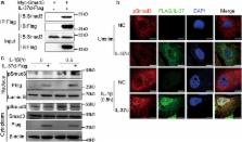
          IL-37 is a new member of IL-1 family and possesses five different isoforms (named as IL-37 a–e). IL-37b has been demonstrated as a physiological suppressor of immune responses. However, the function of other isoforms remains unknown. Here, we show that IL-37d possesses anti-inflammatory roles both in vitro and in vivo. Firstly, IL-37d is expressed in peripheral blood mononuclear cells (PBMCs) and umbilical cords-derived mesenchymal stem cells (UCMSCs). Secondly, IL-37d overexpression markedly inhibits IL-1β-induced IL-6 production in A549 cells. Consistently, bone marrow-derived macrophages (BMDMs) from IL-37d transgenic mice express low levels of pro-inflammatory cytokines (such as IL-6 and TNF-α) following LPS stimulation, compared with those from wild-type mice. Furthermore, IL-37d transgenic mice produce less pro-inflammatory cytokines, and show much less degree of LPS-induced endotoxemia in vivo. Mechanistically, IL-37d interacts with Smad3 and promotes nuclear translocation of pSmad3. SIS3 (a specific Smad3 inhibitor) treatment completely blocks the inhibitory effects of IL-37d. Thus, our data indicate that IL-37d is a functional cytokine that negatively regulates pro-inflammatory cytokines expression in a Smad3-dependent manner.

          Related collections

          Most cited references38

          • Record: found
          • Abstract: found
          • Article: not found

          Interleukin 37 expression protects mice from colitis.

          IL-37, a newly described member of the IL-1 family, functions as a fundamental inhibitor of innate inflammation and immunity. In the present study, we examined a role for IL-37 during experimental colitis. A transgenic mouse strain was generated to express human IL-37 (hIL-37tg), and these mice were subjected to dextran sulfate sodium (DSS)-induced colitis. Despite the presence of a CMV promoter to drive expression of IL-37, mRNA transcripts were not present in colons at the resting state. Expression was observed only upon disruption of the epithelial barrier, with a six- to sevenfold increase (P = 0.02) on days 3 and 5 after continuous exposure to DSS. During the development of colitis, clinical disease scores were reduced by 50% (P < 0.001), and histological indices of colitis were one-third less in hIL-37tg mice compared with WT counterparts (P < 0.001). Reduced inflammation was associated with decreased leukocyte recruitment into the colonic lamina propria. In addition, release of IL-1β and TNFα from ex vivo colonic explant tissue was decreased 5- and 13-fold, respectively, compared with WT (P ≤ 0.005), whereas IL-10 was increased sixfold (P < 0.001). However, IL-10 was not required for the anti-inflammatory effects of IL-37 because IL-10-receptor antibody blockade did not reverse IL-37-mediated protection. Mechanistically, IL-37 originating from hematopoietic cells was sufficient to exert anti-inflammatory effects because WT mice reconstituted with hIL-37tg bone marrow were protected from colitis. Thus, IL-37 emerges as key modulator of intestinal inflammation.
            Bookmark
            • Record: found
            • Abstract: found
            • Article: not found

            Smad3 and Smad4 cooperate with c-Jun/c-Fos to mediate TGF-beta-induced transcription.

            Smad proteins transduce signals for transforming growth factor-beta (TGF-beta)-related factors. Smad proteins activated by receptors for TGF-beta form complexes with Smad4. These complexes are translocated into the nucleus and regulate ligand-induced gene transcription. 12-O-tetradecanoyl-13-acetate (TPA)-responsive gene promoter elements (TREs) are involved in the transcriptional responses of several genes to TGF-beta (refs 5-8). AP-1 transcription factors, composed of c-Jun and c-Fos, bind to and direct transcription from TREs, which are therefore known as AP1-binding sites. Here we show that Smad3 interacts directly with the TRE and that Smad3 and Smad4 can activate TGF-beta-inducible transcription from the TRE in the absence of c-Jun and c-Fos. Smad3 and Smad4 also act together with c-Jun and c-Fos to activate transcription in response to TGF-beta, through a TGF-beta-inducible association of c-Jun with Smad3 and an interaction of Smad3 and c-Fos. These interactions complement interactions between c-Jun and c-Fos, and between Smad3 and Smad4. This mechanism of transcriptional activation by TGF-beta, through functional and physical interactions between Smad3-Smad4 and c-Jun-c-Fos, shows that Smad signalling and MAPK/JNK signalling converge at AP1-binding promoter sites.
              Bookmark
              • Record: found
              • Abstract: found
              • Article: not found

              IL-37: a new anti-inflammatory cytokine of the IL-1 family.

              The IL-1 family of cytokines encompasses eleven proteins that each share a similar β-barrel structure and bind to Ig-like receptors. Some of the IL-1-like cytokines have been well characterised, and play key roles in the development and regulation of inflammation. Indeed, IL-1α (IL-1F1), IL-1β (IL-1F2), and IL-18 (IL-1F4) are well-known inflammatory cytokines active in the initiation of the inflammatory reaction and in driving Th1 and Th17 inflammatory responses. In contrast, IL-1 receptor antagonist (IL-1Ra, IL-1F3) and the receptor antagonist binding to IL-1Rrp2 (IL-36Ra, IL-1F5) reduce inflammation by blocking the binding of the agonist receptor ligands. In the case of IL-37 (IL-1F7), of which five different splice variants have been described, less is known of its function, and identification of the components of a heterodimeric receptor complex remains unclear. Some studies suggest that IL-37 binds to the α chain of the IL-18 receptor in a non-competitive fashion, and this may explain some of the disparate biological effects that have been reported for mice deficient in the IL-18R. The biological properties of IL-37 are mainly those of down-regulating inflammation, as assessed in models where human IL-37 is expressed in mice. In this review, an overview of the role of IL-37 in the regulation of inflammation is presented. The finding that IL-37 also locates to the nucleus, as do IL-1α and IL-33, for receptor-independent organ/tissue-specific regulation of inflammation is also reviewed.
                Bookmark

                Author and article information

                Contributors
                +86 531 88382038 , zhanglining@sdu.edu.cn
                Journal
                Cell Death Dis
                Cell Death Dis
                Cell Death & Disease
                Nature Publishing Group UK (London )
                2041-4889
                22 May 2018
                22 May 2018
                June 2018
                : 9
                : 6
                : 582
                Affiliations
                [1 ]ISNI 0000 0004 1761 1174, GRID grid.27255.37, Department of Immunology and Key Laboratory of Infection and Immunity of Shandong Province, , Shandong University School of Basic Medical Sciences, ; 44 #Wenhua Xi Road, 250012 Jinan, China
                [2 ]ISNI 0000 0004 1761 1174, GRID grid.27255.37, Department of pathogenic biology, , Shandong University School of Basic Medicine Sciences, ; 44# Wenhua Xi Road, 250012 Jinan, China
                [3 ]ISNI 0000 0001 2297 5165, GRID grid.94365.3d, Mucosal Immunology Section, National Institute of Dental and Craniofacial Research (NIDCR), , National Institutes of Health (NIH), ; 30 Convent Drive, Bethesda, Maryland 20892 USA
                Article
                664
                10.1038/s41419-018-0664-0
                5964144
                29789615
                58247c45-9e4c-45af-a8f1-99cba20b8c39
                © The Author(s) 2018

                Open Access This article is licensed under a Creative Commons Attribution 4.0 International License, which permits use, sharing, adaptation, distribution and reproduction in any medium or format, as long as you give appropriate credit to the original author(s) and the source, provide a link to the Creative Commons license, and indicate if changes were made. The images or other third party material in this article are included in the article’s Creative Commons license, unless indicated otherwise in a credit line to the material. If material is not included in the article’s Creative Commons license and your intended use is not permitted by statutory regulation or exceeds the permitted use, you will need to obtain permission directly from the copyright holder. To view a copy of this license, visit http://creativecommons.org/licenses/by/4.0/.

                History
                : 22 December 2017
                : 10 April 2018
                : 3 May 2018
                Categories
                Article
                Custom metadata
                © The Author(s) 2018

                Cell biology
                Cell biology

                Comments

                Comment on this article