25
views
0
recommends
+1 Recommend
0 collections
    0
    shares
      • Record: found
      • Abstract: found
      • Article: found
      Is Open Access

      Characterization of blunt chest trauma in a long-term porcine model of severe multiple trauma

      research-article

      Read this article at

      Bookmark
          There is no author summary for this article yet. Authors can add summaries to their articles on ScienceOpen to make them more accessible to a non-specialist audience.

          Abstract

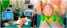
          Chest trauma has a significant relevance on outcome after severe trauma. Clinically, impaired lung function typically occurs within 72 hours after trauma. However, the underlying pathophysiological mechanisms are still not fully elucidated. Therefore, we aimed to establish an experimental long-term model to investigate physiological, morphologic and inflammatory changes, after severe trauma. Male pigs ( sus scrofa) sustained severe trauma (including unilateral chest trauma, femur fracture, liver laceration and hemorrhagic shock). Additionally, non-injured animals served as sham controls. Chest trauma resulted in severe lung damage on both CT and histological analyses. Furthermore, severe inflammation with a systemic increase of IL-6 (p = 0.0305) and a local increase of IL-8 in BAL (p = 0.0009) was observed. The pO 2/FiO 2 ratio in trauma animals decreased over the observation period (p < 0.0001) but not in the sham group (p = 0.2967). Electrical Impedance Tomography (EIT) revealed differences between the traumatized and healthy lung (p < 0.0001). In conclusion, a clinically relevant, long-term model of blunt chest trauma with concomitant injuries has been developed. This reproducible model allows to examine local and systemic consequences of trauma and is valid for investigation of potential diagnostic or therapeutic options. In this context, EIT might represent a radiation-free method for bedside diagnostics.

          Related collections

          Most cited references67

          • Record: found
          • Abstract: found
          • Article: not found

          Effect of whole-body CT during trauma resuscitation on survival: a retrospective, multicentre study.

          The number of trauma centres using whole-body CT for early assessment of primary trauma is increasing. There is no evidence to suggest that use of whole-body CT has any effect on the outcome of patients with major trauma. We therefore compared the probability of survival in patients with blunt trauma who had whole-body CT during resuscitation with those who had not. In a retrospective, multicentre study, we used the data recorded in the trauma registry of the German Trauma Society to calculate the probability of survival according to the trauma and injury severity score (TRISS), revised injury severity classification (RISC) score, and standardised mortality ratio (SMR, ratio of recorded to expected mortality) for 4621 patients with blunt trauma given whole-body or non-whole-body CT. 1494 (32%) of 4621 patients were given whole-body CT. Mean age was 42.6 years (SD 20.7), 3364 (73%) were men, and mean injury-severity score was 29.7 (13.0). SMR based on TRISS was 0.745 (95% CI 0.633-0.859) for patients given whole-body CT versus 1.023 (0.909-1.137) for those given non-whole-body CT (p<0.001). SMR based on the RISC score was 0.865 (0.774-0.956) for patients given whole-body CT versus 1.034 (0.959-1.109) for those given non-whole-body CT (p=0.017). The relative reduction in mortality based on TRISS was 25% (14-37) versus 13% (4-23) based on RISC score. Multivariate adjustment for hospital level, year of trauma, and potential centre effects confirmed that whole-body CT is an independent predictor for survival (p
            Bookmark
            • Record: found
            • Abstract: found
            • Article: not found

            Assessment of regional lung recruitment and derecruitment during a PEEP trial based on electrical impedance tomography.

            To investigate whether electrical impedance tomography (EIT) is capable of monitoring regional lung recruitment and lung collapse during a positive end-expiratory pressure (PEEP) trial. Experimental animal study of acute lung injury. Six pigs with saline-lavage-induced acute lung injury. An incremental and decremental PEEP trial at ten pressure levels was performed. Ventilatory, gas exchange, and hemodynamic parameters were automatically recorded. EIT and computed tomography (CT) scans of the same slice were simultaneously taken at each PEEP level. A significant correlation between EIT and CT analyses of end-expiratory gas volumes (r=0.98 up to 0.99) and tidal volumes (r=0.55 up to r=0.88) could be demonstrated. Changes in global and regional tidal volumes and arterial oxygenation (PaO2/FiO2) demonstrated recruitment/derecruitment during the trial, but at different onsets. During the decremental trial, derecruitment first occurred in dependent lung areas. This was indicated by lowered regional tidal volumes measured in this area and by a decrease of PaO2/FiO2. At the same time, the global tidal volume still continued to increase, because the increase of ventilation of the non-dependent areas was higher than the loss in the dependent areas. This indicates that opposing regional changes might cancel each other out when combined in a global parameter. EIT is suitable for monitoring the dynamic effects of PEEP variations on the regional change of tidal volume. It is superior to global ventilation parameters in assessing the beginning of alveolar recruitment and lung collapse.
              Bookmark
              • Record: found
              • Abstract: found
              • Article: not found

              Appraisal of early evaluation of blunt chest trauma: development of a standardized scoring system for initial clinical decision making.

              Current techniques for assessment of chest trauma rely on clinical diagnoses or scoring systems. However, there is no generally accepted standard for early judgement of the severity of these injuries, especially in regards to related complications. This drawback may have a significant impact on the management of skeletal injuries, which are frequently associated with chest trauma. However, no convincing conclusions can be determined until standardization of the degrees of chest trauma is achieved. We investigated the role of early clinical and radiologic assessment techniques on outcome in patients with blunt multiple trauma and thoracic injuries and developed a new scoring system for early evaluation of chest trauma. A retrospective investigation was performed on the basis of 4,571 blunt polytrauma (Injury Severity Score [ISS] > or = 18) patients admitted to our unit. Inclusion criteria were treatment of thoracic injury that required intensive care therapy, initial Glasgow Coma Scale score greater than 8 points, and no local or systemic infection. Patients with thoracic trauma and multiple associated injuries (ISS > or = 18) were included. In all patients, the association between various parameters of the thoracic injuries and subsequent mortality and morbidity was investigated. A total of 1,495 patients fulfilled the inclusion criteria. Patients' medical records and chest radiographs were reevaluated between May 1, 1998, and June 1, 1999. The association between rib fractures and chest-related death was low (> three ribs unilateral, mortality 17.3%, odds ratio 1.01) unless bilateral involvement was present (> three ribs bilateral, mortality 40.9%, odds ratio 3.43). Injuries to the lung parenchyma, as determined by plain radiography, were associated with chest-related death, especially if the injuries were bilateral or associated with hemopneumothorax (lung contusion unilateral, mortality 25.2%, odds ratio 1.82; lung contusion bilateral + hemopneumothorax, mortality 53.3%, odds ratio 5.1). When plain anteroposterior chest radiographs were used, the diagnostic rate of rib fractures (< or = three ribs) increased slightly, from 77.1% to 97.3% during the first 24 hours of admission. In contrast, pulmonary contusions were often not diagnosed until 24 hours after admission (47.3% at admission, 92.4% at 24 h, p = 0.002). A new composite scoring system (thoracic trauma severity score) was developed that combines several variables: injuries to the chest wall, intrathoracic lesions, injuries involving the pleura, admission PaO2/FIO2 ratio, and patient age. The receiver operating characteristic curve demonstrated an adequate discrimination, as demonstrated by a value of 0.924 for the development set and 0.916 for the validation set. The score was also superior to the ISS (0.881) or the thorax Abbreviated Injury Score (0.693). Radiographically determined injuries to the lung parenchyma have a closer association with adverse outcome than chest-wall injuries but are often not diagnosed until 24 hours after injury. Therefore, clinical decision making, such as about the choice of surgery for long bone fractures, may be flawed if this information is used alone. A new thoracic trauma severity score may serve as an additional tool to improve the accuracy of the prediction of thoracic trauma-related complications.
                Bookmark

                Author and article information

                Journal
                Sci Rep
                Sci Rep
                Scientific Reports
                Nature Publishing Group
                2045-2322
                21 December 2016
                2016
                : 6
                : 39659
                Affiliations
                [1 ]Department of Orthopaedic Trauma, RWTH Aachen University , Germany
                [2 ]Harald Tscherne Research Laboratory, RWTH Aachen University , Germany
                [3 ]Department of Intensive Care and Intermediate Care, RWTH Aachen University , Germany
                [4 ]Chair for Medical Information Technology, Helmholtz-Institute for Biomedical Engineering, RWTH Aachen University , Aachen, Germany
                [5 ]Department of Medical Statistics, RWTH Aachen University , Germany
                [6 ]Medical School, Sigmund Freud Private University , Vienna, Austria
                [7 ]Department of Trauma-, Hand- and Reconstructive Surgery, University of Frankfurt/Main , Germany
                [8 ]Experimental Trauma Surgery, Department of Trauma Surgery, Klinikum rechts der Isar, Technical University of Munich , Germany
                [9 ]Department of Orthopedic Trauma, Hand-, Plastic-, and Reconstructive Surgery, University of Ulm , Germany
                [10 ]Institute for Laboratory Animal Science and Experimental Surgery, RWTH Aachen University , Germany
                [11 ]Institute of Pharmacology and Toxicology, RWTH Aachen University , Germany
                Author notes
                [*]

                These authors contributed equally to this work.

                Article
                srep39659
                10.1038/srep39659
                5175194
                28000769
                5f224679-85f7-472d-8c19-54647cb541bc
                Copyright © 2016, The Author(s)

                This work is licensed under a Creative Commons Attribution 4.0 International License. The images or other third party material in this article are included in the article’s Creative Commons license, unless indicated otherwise in the credit line; if the material is not included under the Creative Commons license, users will need to obtain permission from the license holder to reproduce the material. To view a copy of this license, visit http://creativecommons.org/licenses/by/4.0/

                History
                : 04 October 2016
                : 24 November 2016
                Categories
                Article

                Uncategorized
                Uncategorized

                Comments

                Comment on this article