10
views
0
recommends
+1 Recommend
0 collections
    0
    shares
      • Record: found
      • Abstract: found
      • Article: found
      Is Open Access

      Kinesin-5 inhibitor resistance is driven by kinesin-12

      research-article

      Read this article at

      Bookmark
          There is no author summary for this article yet. Authors can add summaries to their articles on ScienceOpen to make them more accessible to a non-specialist audience.

          Abstract

          The kinesin-5 Eg5 is essential for mitotic progression, and the lethal effects of Eg5 inhibitors make these inhibitors attractive candidates for chemotherapy drugs. Sturgill et al. show that kinesin-12 and a nonmotile Eg5 mutant form an alternative spindle assembly pathway that provides resistance to Eg5 inhibitors.

          Abstract

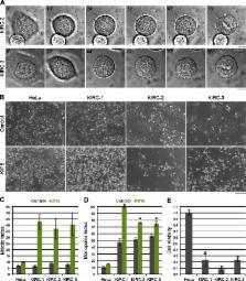
          The microtubule (MT) cytoskeleton bipolarizes at the onset of mitosis to form the spindle. In animal cells, the kinesin-5 Eg5 primarily drives this reorganization by actively sliding MTs apart. Its primacy during spindle assembly renders Eg5 essential for mitotic progression, demonstrated by the lethal effects of kinesin-5/Eg5 inhibitors (K5Is) administered in cell culture. However, cultured cells can acquire resistance to K5Is, indicative of alternative spindle assembly mechanisms and/or pharmacological failure. Through characterization of novel K5I-resistant cell lines, we unveil an Eg5 motility-independent spindle assembly pathway that involves both an Eg5 rigor mutant and the kinesin-12 Kif15. This pathway centers on spindle MT bundling instead of Kif15 overexpression, distinguishing it from those previously described. We further show that large populations (∼10 7 cells) of HeLa cells require Kif15 to survive K5I treatment. Overall, this study provides insight into the functional plasticity of mitotic kinesins during spindle assembly and has important implications for the development of antimitotic regimens that target this process.

          Related collections

          Most cited references52

          • Record: found
          • Abstract: found
          • Article: not found

          Nonsense-mediated mRNA decay: an intricate machinery that shapes transcriptomes.

          Nonsense-mediated mRNA decay (NMD) is probably the best characterized eukaryotic RNA degradation pathway. Through intricate steps, a set of NMD factors recognize and degrade mRNAs with translation termination codons that are positioned in abnormal contexts. However, NMD is not only part of a general cellular quality control system that prevents the production of aberrant proteins. Mammalian cells also depend on NMD to dynamically adjust their transcriptomes and their proteomes to varying physiological conditions. In this Review, we discuss how NMD targets mRNAs, the types of mRNAs that are targeted, and the roles of NMD in cellular stress, differentiation and maturation processes.
            Bookmark
            • Record: found
            • Abstract: found
            • Article: not found

            Selective small-molecule inhibitor reveals critical mitotic functions of human CDK1.

            CDK1 is a nonredundant cyclin-dependent kinase (CDK) with an essential role in mitosis, but its multiple functions still are poorly understood at a molecular level. Here we identify a selective small-molecule inhibitor of CDK1 that reversibly arrests human cells at the G(2)/M border of the cell cycle and allows for effective cell synchronization in early mitosis. Inhibition of CDK1 during cell division revealed that its activity is necessary and sufficient for maintaining the mitotic state of the cells, preventing replication origin licensing and premature cytokinesis. Although CDK1 inhibition for up to 24 h is well tolerated, longer exposure to the inhibitor induces apoptosis in tumor cells, suggesting that selective CDK1 inhibitors may have utility in cancer therapy.
              Bookmark
              • Record: found
              • Abstract: found
              • Article: not found

              The bipolar mitotic kinesin Eg5 moves on both microtubules that it crosslinks.

              During cell division, mitotic spindles are assembled by microtubule-based motor proteins. The bipolar organization of spindles is essential for proper segregation of chromosomes, and requires plus-end-directed homotetrameric motor proteins of the widely conserved kinesin-5 (BimC) family. Hypotheses for bipolar spindle formation include the 'push-pull mitotic muscle' model, in which kinesin-5 and opposing motor proteins act between overlapping microtubules. However, the precise roles of kinesin-5 during this process are unknown. Here we show that the vertebrate kinesin-5 Eg5 drives the sliding of microtubules depending on their relative orientation. We found in controlled in vitro assays that Eg5 has the remarkable capability of simultaneously moving at approximately 20 nm s(-1) towards the plus-ends of each of the two microtubules it crosslinks. For anti-parallel microtubules, this results in relative sliding at approximately 40 nm s(-1), comparable to spindle pole separation rates in vivo. Furthermore, we found that Eg5 can tether microtubule plus-ends, suggesting an additional microtubule-binding mode for Eg5. Our results demonstrate how members of the kinesin-5 family are likely to function in mitosis, pushing apart interpolar microtubules as well as recruiting microtubules into bundles that are subsequently polarized by relative sliding.
                Bookmark

                Author and article information

                Journal
                J Cell Biol
                J. Cell Biol
                jcb
                jcb
                The Journal of Cell Biology
                The Rockefeller University Press
                0021-9525
                1540-8140
                25 April 2016
                : 213
                : 2
                : 213-227
                Affiliations
                [1 ]Department of Cell and Developmental Biology, Vanderbilt University Medical Center, Nashville, TN 37232
                [2 ]Department of Cancer Biology, Vanderbilt University Medical Center, Nashville, TN 37232
                Author notes
                Correspondence to Ryoma Ohi: ryoma.ohi@ 123456vanderbilt.edu
                Article
                201507036
                10.1083/jcb.201507036
                5084272
                27091450
                5f5ca3cc-40e0-4357-a66a-cf53156f7dbf
                © 2016 Sturgill et al.

                This article is distributed under the terms of an Attribution–Noncommercial–Share Alike–No Mirror Sites license for the first six months after the publication date (see http://www.rupress.org/terms). After six months it is available under a Creative Commons License (Attribution–Noncommercial–Share Alike 3.0 Unported license, as described at http://creativecommons.org/licenses/by-nc-sa/3.0/).

                History
                : 08 July 2015
                : 10 March 2016
                Funding
                Funded by: National Institutes of Health http://dx.doi.org/10.13039/100000002
                Award ID: R01GM086610
                Categories
                Research Articles
                Article

                Cell biology
                Cell biology

                Comments

                Comment on this article