19
views
0
recommends
+1 Recommend
0 collections
    0
    shares
      • Record: found
      • Abstract: found
      • Article: found
      Is Open Access

      BRE modulates granulosa cell death to affect ovarian follicle development and atresia in the mouse

      research-article

      Read this article at

      Bookmark
          There is no author summary for this article yet. Authors can add summaries to their articles on ScienceOpen to make them more accessible to a non-specialist audience.

          Abstract

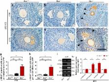
          The BRE (brain and reproductive expression) gene, highly expressed in nervous and reproductive system organs, plays an important role in modulating DNA damage repair under stress response and pathological conditions. Folliculogenesis, a process that ovarian follicle develops into maturation, is closely associated with the interaction between somatic granulosa cell and oocyte. However, the regulatory role of BRE in follicular development remains undetermined. In this context, we found that BRE is normally expressed in the oocytes and granulosa cells from the primordial follicle stage. There was a reduction in follicles number of BRE mutant (BRE −/−) mice. It was attributed to increase the follicular atresia in ovaries, as a result of retarded follicular development. We established that cell proliferation was inhibited, while apoptosis was markedly increased in the granulosa cells in the absence of BRE. In addition, expressions of γ-H2AX (marker for showing DNA double-strand breaks) and DNA damage-relevant genes are both upregulated in BRE −/− mice. In sum, these results suggest that the absence of BRE, deficiency in DNA damage repair, causes increased apoptosis in granulosa cells, which in turn induces follicular atresia in BRE −/− mice.

          Related collections

          Most cited references25

          • Record: found
          • Abstract: found
          • Article: not found

          Cell cycle regulation and neural differentiation.

          The general mechanisms that control the cell cycle in mammalian cells have been studied in depth and several proteins that are involved in the tight regulation of cell cycle progression have been identified. However, the analysis of which molecules participate in cell cycle exit of specific cell lineages is not exhaustive yet. Moreover, the strict relation between cell cycle exit and induction of differentiation has not been fully understood and seems to depend on the cell type. Several in vivo and in vitro studies have been performed in the last few years to address these issues in cells of the nervous system. In this review, we focus our attention on cyclin-cyclin-dependent kinase complexes, cyclin kinase inhibitors, genes of the retinoblastoma family, p53 and N-Myc, and we aim to summarize the latest evidence indicating their involvement in the control of the cell cycle and induction of differentiation in different cell types of the peripheral and central nervous systems. Studies on nervous system tumors and a possible contributory role in tumorigenesis of polyomavirus T antigen are reported to point out the critical contribution of some cell cycle regulators to normal neural and glial development.
            Bookmark
            • Record: found
            • Abstract: found
            • Article: not found

            Premature ovarian failure in androgen receptor-deficient mice.

            Premature ovarian failure (POF) syndrome, an early decline of ovarian function in women, is frequently associated with X chromosome abnormalities ranging from various Xq deletions to complete loss of one of the X chromosomes. However, the genetic locus responsible for the POF remains unknown, and no candidate gene has been identified. Using the Cre/LoxP system, we have disrupted the mouse X chromosome androgen receptor (Ar) gene. Female AR(-/-) mice appeared normal but developed the POF phenotype with aberrant ovarian gene expression. Eight-week-old female AR(-/-) mice are fertile, but they have lower follicle numbers and impaired mammary development, and they produce only half of the normal number of pups per litter. Forty-week-old AR(-/-) mice are infertile because of complete loss of follicles. Genome-wide microarray analysis of mRNA from AR(-/-) ovaries revealed that a number of major regulators of folliculogenesis were under transcriptional control by AR. Our findings suggest that AR function is required for normal female reproduction, particularly folliculogenesis, and that AR is a potential therapeutic target in POF syndrome.
              Bookmark
              • Record: found
              • Abstract: found
              • Article: not found

              Apoptosis in granulosa cells during follicular atresia: relationship with steroids and insulin-like growth factors.

              It is well known that during mammalian ovarian follicular development, the majority of follicles undergo atresia at various stages of their development. However, the mechanisms controlling this selection process remain unknown. In this study, we investigated apoptosis in granulosa cells during goat follicular atresia by terminal deoxynucleotidyl transferase-mediated dUTP nick end labeling (TUNEL). The changes in the levels of steroids, insulin-like growth factors (IGFs) and IGF receptors were studied by radioimmunoassay (RIA) and semi-quantitative reverse transcription-PCR. We found that the percentage of apoptotic granulosa cells in the atretic (A) follicles was significantly higher than that in the slightly atretic (SA) and healthy (H) follicles. The level of estradiol and the ratio of estradiol to progesterone in H follicles were significantly higher than those in A follicles. On the other hand, the level of progesterone was not significantly different among these follicle types. We also found that the level of IGF-I in H follicles was higher than in SA and A follicles, whereas the amount of IGF-II did not vary significantly. The expression of IGF receptor also decreased in A follicles as compared to that in H and SA follicles. These results suggested that estradiol and IGF-I might be involved in controlling apoptosis in granulosa cells during follicular atresia.
                Bookmark

                Author and article information

                Journal
                Cell Death Dis
                Cell Death Dis
                Cell Death & Disease
                Nature Publishing Group
                2041-4889
                March 2017
                23 March 2017
                1 March 2017
                : 8
                : 3
                : e2697
                Affiliations
                [1 ]Division of Histology and Embryology, Key Laboratory for Regenerative Medicine of the Ministry of Education, Medical College, Jinan University , Guangzhou 510632, China
                [2 ]Chinese Medicine College, Jinan University , Guangzhou 510632, China
                [3 ]Key Laboratory for Regenerative Medicine of the Ministry of Education, School of Biomedical Sciences, Chinese University of Hong Kong , Shatin, Hong Kong
                [4 ]Division of Cell and Developmental Biology, University of Dundee , Dundee DD1 5EH, UK
                Author notes
                [* ]Division of Histology and Embryology, Key Laboratory for Regenerative Medicine of the Ministry of Education, Medical College, Jinan University , Guangzhou 510632, China. Tel: +86 020 8522 8316; Fax: +86 020 8522 1343; E-mail: yang_xuesong@ 123456126.com
                [* ]Key Laboratory for Regenerative Medicine of the Ministry of Education, School of Biomedical Sciences, Chinese University of Hong Kong , Shatin, Hong Kong. Tel: (852) 3943 6785; Fax: (852) 2603 7902; E-mail: kaholee@ 123456cuhk.edu.hk
                [5]

                These authors contributed equally to this work.

                Article
                cddis201791
                10.1038/cddis.2017.91
                5386581
                28333135
                694b9f82-d157-4d30-a661-5e1dbfbe35f5
                Copyright © 2017 The Author(s)

                Cell Death and Disease is an open-access journal published by Nature Publishing Group. This work is licensed under a Creative Commons Attribution 4.0 International License. The images or other third party material in this article are included in the article's Creative Commons license, unless indicated otherwise in the credit line; if the material is not included under the Creative Commons license, users will need to obtain permission from the license holder to reproduce the material. To view a copy of this license, visit http://creativecommons.org/licenses/by/4.0/

                History
                : 05 October 2016
                : 09 January 2017
                : 09 February 2017
                Categories
                Original Article

                Cell biology
                Cell biology

                Comments

                Comment on this article