84
views
0
recommends
+1 Recommend
0 collections
    1
    shares
      • Record: found
      • Abstract: found
      • Article: found
      Is Open Access

      Regulation of Adaptive Immunity; The Role of Interleukin-10

      review-article

      Read this article at

      Bookmark
          There is no author summary for this article yet. Authors can add summaries to their articles on ScienceOpen to make them more accessible to a non-specialist audience.

          Abstract

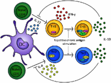
          Since the discovery of interleukin-10 (IL-10) in the 1980s, a large body of work has led to its recognition as a pleiotropic immunomodulatory cytokine that affects both the innate and adaptive immune systems. IL-10 is produced by a wide range of cell types, but for the purposes of this review we shall focus on IL-10 secreted by CD4 + T cells. Here we describe the importance of IL-10 as a mediator of suppression used by both FoxP3 + and FoxP3 T regulatory cells. Moreover, we discuss the molecular events leading to the induction of IL-10 secretion in T helper cell subsets, where it acts as a pivotal negative feedback mechanism. Finally we discuss how a greater understanding of this principle has allowed for the design of more efficient, antigen-specific immunotherapy strategies to exploit this natural phenomenon clinically.

          Related collections

          Most cited references144

          • Record: found
          • Abstract: found
          • Article: not found

          Two types of mouse T helper cell. IV. Th2 clones secrete a factor that inhibits cytokine production by Th1 clones

          A cytokine synthesis inhibitory factor (CSIF) is secreted by Th2 clones in response to Con A or antigen stimulation, but is absent in supernatants from Con A-induced Th1 clones. CSIF can inhibit the production of IL-2, IL-3, lymphotoxin (LT)/TNF, IFN-gamma, and granulocyte-macrophage CSF (GM-CSF) by Th1 cells responding to antigen and APC, but Th2 cytokine synthesis is not significantly affected. Transforming growth factor beta (TGF-beta) also inhibits IFN-gamma production, although less effectively than CSIF, whereas IL-2 and IL-4 partially antagonize the activity of CSIF. CSIF inhibition of cytokine synthesis is not complete, since early cytokine synthesis (before 8 h) is not significantly affected, whereas later synthesis is strongly inhibited. In the presence of CSIF, IFN-gamma mRNA levels are reduced slightly at 8, and strongly at 12 h after stimulation. Inhibition of cytokine expression by CSIF is not due to a general reduction in Th1 cell viability, since actin mRNA levels were not reduced, and proliferation of antigen-stimulated cells in response to IL-2, was unaffected. Biochemical characterization, mAbs, and recombinant or purified cytokines showed that CSIF is distinct from IL-1, IL-2, IL-3, IL-4, IL-5, IL-6, IL-7, IFN-gamma, GM-CSF, TGF-beta, TNF, LT, and P40. The potential role of CSIF in crossregulation of Th1 and Th2 responses is discussed.
            Bookmark
            • Record: found
            • Abstract: found
            • Article: not found

            The interleukin 23 receptor is essential for the terminal differentiation of interleukin 17-producing effector T helper cells in vivo.

            Interleukin 23 (IL-23) is required for autoimmune inflammation mediated by IL-17-producing helper T cells (T(H)-17 cells) and has been linked to many human immune disorders. Here we restricted deficiency in the IL-23 receptor to defined cell populations in vivo to investigate the requirement for IL-23 signaling in the development and function of T(H)-17 cells in autoimmunity, inflammation and infection. In the absence of IL-23, T(H)-17 development was stalled at the early activation stage. T(H)-17 cells failed to downregulate IL-2 and also failed to maintain IL-17 production or upregulate expression of the IL-7 receptor alpha-chain. These defects were associated with less proliferation; consequently, fewer effector T(H)-17 cells were produced in the lymph nodes and hence available to emigrate to the bloodstream and tissues.
              Bookmark
              • Record: found
              • Abstract: found
              • Article: not found

              Predominant TH2-like bronchoalveolar T-lymphocyte population in atopic asthma.

              In atopic asthma, activated T helper lymphocytes are present in bronchial-biopsy specimens and bronchoalveolar-lavage (BAL) fluid, and their production of cytokines may be important in the pathogenesis of this disorder. Different patterns of cytokine release are characteristic of certain subgroups of T helper cells, termed TH1 and TH2, the former mediating delayed-type hypersensitivity and the latter mediating IgE synthesis and eosinophilia. The pattern of cytokine production in atopic asthma is unknown. We assessed cells obtained by BAL in subjects with mild atopic asthma and in normal control subjects for the expression of messenger RNA (mRNA) for interleukin-2, 3, 4, and 5, granulocyte-macrophage colony-stimulating factor (GM-CSF), and interferon gamma by in situ hybridization with 32P-labeled complementary RNA. Localization of mRNA to BAL T cells was assessed by simultaneous in situ hybridization and immunofluorescence and by in situ hybridization after immunomagnetic enrichment or depletion of T cells. As compared with the control subjects, the subjects with asthma had more BAL cells per 1000 cell that were positive for mRNA for interleukin-2 (P less than 0.05), 3 (P less than 0.01), 4 (P less than 0.001), and 5 (P less than 0.001) and GM-CSF (P less than 0.001). There was no significant difference between the two groups in the number of cells expressing mRNA for interferon gamma. In the subjects with asthma, mRNA for interleukin-4 and 5 was expressed predominantly by T lymphocytes. Atopic asthma is associated with activation in the bronchi of the interleukin-3, 4, and 5 and GM-CSF gene cluster, a pattern compatible with predominant activation of the TH2-like T-cell population.
                Bookmark

                Author and article information

                Journal
                Front Immunol
                Front Immunol
                Front. Immunol.
                Frontiers in Immunology
                Frontiers Media S.A.
                1664-3224
                13 May 2013
                31 May 2013
                2013
                : 4
                : 129
                Affiliations
                [1]School of Cellular and Molecular Medicine, University of Bristol , Bristol, UK
                Author notes

                Edited by: Eyad Elkord, United Arab Emirates University, UAE; University of Salford and University of Manchester, UK

                Reviewed by: Giovanna Lombardi, Kings College London, UK; Bin Li, Chinese Academy of Sciences, China

                *Correspondence: David C. Wraith, School of Cellular and Molecular Medicine, Medical Sciences Building, University Walk, Bristol BS8 1TD, UK e-mail: d.c.wraith@ 123456bristol.ac.uk

                This article was submitted to Frontiers in Immunological Tolerance, a specialty of Frontiers in Immunology.

                Article
                10.3389/fimmu.2013.00129
                3668291
                23755052
                697a226a-6659-479e-92bc-da57f354e859
                Copyright © 2013 Ng, Britton, Hill, Verhagen, Burton and Wraith.

                This is an open-access article distributed under the terms of the Creative Commons Attribution License, which permits use, distribution and reproduction in other forums, provided the original authors and source are credited and subject to any copyright notices concerning any third-party graphics etc.

                History
                : 01 May 2013
                : 15 May 2013
                Page count
                Figures: 3, Tables: 0, Equations: 0, References: 179, Pages: 13, Words: 12777
                Categories
                Immunology
                Review Article

                Immunology
                allergy,autoimmunity,cytokines,immune regulation,immunotherapy,interleukin-10,regulatory t cells,t helper cells

                Comments

                Comment on this article