5
views
0
recommends
+1 Recommend
0 collections
    0
    shares
      • Record: found
      • Abstract: found
      • Article: found
      Is Open Access

      miR-143 promotes angiogenesis and osteoblast differentiation by targeting HDAC7

      research-article

      Read this article at

      Bookmark
          There is no author summary for this article yet. Authors can add summaries to their articles on ScienceOpen to make them more accessible to a non-specialist audience.

          Abstract

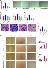
          The regulation of bone formation and detailed mechanisms are still largely elusive, and the roles of microRNAs in this process have attracted much attention. Recently, a specific subtype of CD31 hiendomucin hi (CD31 hiEMCN hi) endothelium has been identified to promote bone formation, together with osteoblast development. However, the role of microRNA143 in the generation of CD31 hi EMCN hi endothelium and bone formation remains unknown. In this study, we found that miR-143 was expressed both in osteoblast cells and CD31 hiEMCN hi endothelial cells. Serum miR-143 level was negatively correlated with age in humans. Overexpression of miR-143 promoted osteoblast formation and angiogenic effects. Furthermore, CD31 hiEmcn hi vessels and osteoblast formation were significantly inhibited in miR-143 knockout mice. Mechanistically, inhibitor HDAC7 was directly targeted by miR-143 and knockdown of HDAC7 was found to rescue the function of miR-143 deficiency. Thus, miR-143 promotes angiogenesis coupling with osteoblast differentiation by targeting HDAC7, which may serve as a potential target in angiogenic and osteogenic diseases.

          Related collections

          Most cited references29

          • Record: found
          • Abstract: found
          • Article: not found

          Coupling of angiogenesis and osteogenesis by a specific vessel subtype in bone.

          The mammalian skeletal system harbours a hierarchical system of mesenchymal stem cells, osteoprogenitors and osteoblasts sustaining lifelong bone formation. Osteogenesis is indispensable for the homeostatic renewal of bone as well as regenerative fracture healing, but these processes frequently decline in ageing organisms, leading to loss of bone mass and increased fracture incidence. Evidence indicates that the growth of blood vessels in bone and osteogenesis are coupled, but relatively little is known about the underlying cellular and molecular mechanisms. Here we identify a new capillary subtype in the murine skeletal system with distinct morphological, molecular and functional properties. These vessels are found in specific locations, mediate growth of the bone vasculature, generate distinct metabolic and molecular microenvironments, maintain perivascular osteoprogenitors and couple angiogenesis to osteogenesis. The abundance of these vessels and associated osteoprogenitors was strongly reduced in bone from aged animals, and pharmacological reversal of this decline allowed the restoration of bone mass.
            Bookmark
            • Record: found
            • Abstract: found
            • Article: not found

            Endothelial Notch activity promotes angiogenesis and osteogenesis in bone.

            Blood vessel growth in the skeletal system and osteogenesis seem to be coupled, suggesting the existence of molecular crosstalk between endothelial and osteoblastic cells. Understanding the nature of the mechanisms linking angiogenesis and bone formation should be of great relevance for improved fracture healing or prevention of bone mass loss. Here we show that vascular growth in bone involves a specialized, tissue-specific form of angiogenesis. Notch signalling promotes endothelial cell proliferation and vessel growth in postnatal long bone, which is the opposite of the well-established function of Notch and its ligand Dll4 in the endothelium of other organs and tumours. Endothelial-cell-specific and inducible genetic disruption of Notch signalling in mice not only impaired bone vessel morphology and growth, but also led to reduced osteogenesis, shortening of long bones, chondrocyte defects, loss of trabeculae and decreased bone mass. On the basis of a series of genetic experiments, we conclude that skeletal defects in these mutants involved defective angiocrine release of Noggin from endothelial cells, which is positively regulated by Notch. Administration of recombinant Noggin, a secreted antagonist of bone morphogenetic proteins, restored bone growth and mineralization, chondrocyte maturation, the formation of trabeculae and osteoprogenitor numbers in endothelial-cell-specific Notch pathway mutants. These findings establish a molecular framework coupling angiogenesis, angiocrine signals and osteogenesis, which may prove significant for the development of future therapeutic applications.
              Bookmark
              • Record: found
              • Abstract: found
              • Article: not found

              MicroRNA-188 regulates age-related switch between osteoblast and adipocyte differentiation.

              Bone marrow mesenchymal stem cells (BMSCs) exhibit an age-dependent reduction in osteogenesis that is accompanied by an increased propensity toward adipocyte differentiation. This switch increases adipocyte numbers and decreases the number of osteoblasts, contributing to age-related bone loss. Here, we found that the level of microRNA-188 (miR-188) is markedly higher in BMSCs from aged compared with young mice and humans. Compared with control mice, animals lacking miR-188 showed a substantial reduction of age-associated bone loss and fat accumulation in bone marrow. Conversely, mice with transgenic overexpression of miR-188 in osterix+ osteoprogenitors had greater age-associated bone loss and fat accumulation in bone marrow relative to WT mice. Moreover, using an aptamer delivery system, we found that BMSC-specific overexpression of miR-188 in mice reduced bone formation and increased bone marrow fat accumulation. We identified histone deacetylase 9 (HDAC9) and RPTOR-independent companion of MTOR complex 2 (RICTOR) as the direct targets of miR-188. Notably, BMSC-specific inhibition of miR-188 by intra-bone marrow injection of aptamer-antagomiR-188 increased bone formation and decreased bone marrow fat accumulation in aged mice. Together, our results indicate that miR-188 is a key regulator of the age-related switch between osteogenesis and adipogenesis of BMSCs and may represent a potential therapeutic target for age-related bone loss.
                Bookmark

                Author and article information

                Contributors
                doctorjif@126.com
                tanghao1978@163.com
                Journal
                Cell Death Dis
                Cell Death Dis
                Cell Death & Disease
                Nature Publishing Group UK (London )
                2041-4889
                9 March 2020
                9 March 2020
                March 2020
                : 11
                : 3
                : 179
                Affiliations
                [1 ]ISNI 0000 0004 0369 1660, GRID grid.73113.37, Department of Orthopedics, Changhai Hospital, , Second Military Medical University, ; Shanghai, China
                [2 ]Guangdong Key Lab of Orthopedic Technology and Implant Materials, Key Laboratory of Trauma and Tissue Repair of Tropical Area of PLA, Hospital of Orthopedics, General Hospital of Southern Theater Command of PLA, 111 Liuhua Road, Guangzhou, Guangdong 510010 China
                Article
                2377
                10.1038/s41419-020-2377-4
                7062786
                32152265
                698ae038-e7b9-48dc-b76a-6132b3fa32ec
                © The Author(s) 2020

                Open Access This article is licensed under a Creative Commons Attribution 4.0 International License, which permits use, sharing, adaptation, distribution and reproduction in any medium or format, as long as you give appropriate credit to the original author(s) and the source, provide a link to the Creative Commons license, and indicate if changes were made. The images or other third party material in this article are included in the article’s Creative Commons license, unless indicated otherwise in a credit line to the material. If material is not included in the article’s Creative Commons license and your intended use is not permitted by statutory regulation or exceeds the permitted use, you will need to obtain permission directly from the copyright holder. To view a copy of this license, visit http://creativecommons.org/licenses/by/4.0/.

                History
                : 27 June 2019
                : 25 February 2020
                : 26 February 2020
                Funding
                Funded by: FundRef https://doi.org/10.13039/501100001809, National Natural Science Foundation of China (National Science Foundation of China);
                Award ID: 81572637
                Award ID: 81202122
                Award ID: 81702666
                Award ID: 81272942
                Award ID: 30973019
                Award Recipient :
                Categories
                Article
                Custom metadata
                © The Author(s) 2020

                Cell biology
                cell lineage,osteoporosis
                Cell biology
                cell lineage, osteoporosis

                Comments

                Comment on this article