Inviting an author to review:
Find an author and click ‘Invite to review selected article’ near their name.
Search for authorsSearch for similar articles
0
views
0
recommends
+1 Recommend
0 collections
    0
    shares
      • Record: found
      • Abstract: found
      • Article: found
      Is Open Access

      A Machine Learning Model Based on PET/CT Radiomics and Clinical Characteristics Predicts Tumor Immune Profiles in Non-Small Cell Lung Cancer: A Retrospective Multicohort Study

      research-article

      Read this article at

      Bookmark
          There is no author summary for this article yet. Authors can add summaries to their articles on ScienceOpen to make them more accessible to a non-specialist audience.

          Abstract

          Background

          The tumor immune microenvironment (TIME) phenotypes have been reported to mainly impact the efficacy of immunotherapy. Given the increasing use of immunotherapy in cancers, knowing an individual’s TIME phenotypes could be helpful in screening patients who are more likely to respond to immunotherapy. Our study intended to establish, validate, and apply a machine learning model to predict TIME profiles in non-small cell lung cancer (NSCLC) by using 18F-FDG PET/CT radiomics and clinical characteristics.

          Methods

          The RNA-seq data of 1145 NSCLC patients from The Cancer Genome Atlas (TCGA) cohort were analyzed. Then, 221 NSCLC patients from Daping Hospital (DPH) cohort received 18F-FDG PET/CT scans before treatment and CD8 expression of the tumor samples were tested. The Artificial Intelligence Kit software was used to extract radiomic features of PET/CT images and develop a radiomics signature. The models were established by radiomics, clinical features, and radiomics-clinical combination, respectively, the performance of which was calculated by receiver operating curves (ROCs) and compared by DeLong test. Moreover, based on radiomics score (Rad-score) and clinical features, a nomogram was established. Finally, we applied the combined model to evaluate TIME phenotypes of NSCLC patients in The Cancer Imaging Archive (TCIA) cohort (n = 39).

          Results

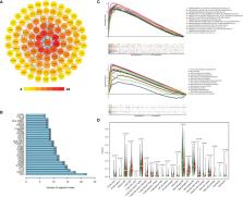
          TCGA data showed CD8 expression could represent the TIME profiles in NSCLC. In DPH cohort, PET/CT radiomics model outperformed CT model (AUC: 0.907 vs. 0.861, P = 0.0314) to predict CD8 expression. Further, PET/CT radiomics-clinical combined model (AUC = 0.932) outperformed PET/CT radiomics model (AUC = 0.907, P = 0.0326) or clinical model (AUC = 0.868, P = 0.0036) to predict CD8 expression. In the TCIA cohort, the predicted CD8-high group had significantly higher immune scores and more activated immune pathways than the predicted CD8-low group ( P = 0.0421).

          Conclusion

          Our study indicates that 18F-FDG PET/CT radiomics-clinical combined model could be a clinically practical method to non-invasively detect the tumor immune status in NSCLCs.

          Related collections

          Most cited references48

          • Record: found
          • Abstract: found
          • Article: not found

          Pembrolizumab versus Chemotherapy for PD-L1–Positive Non–Small-Cell Lung Cancer

          Pembrolizumab is a humanized monoclonal antibody against programmed death 1 (PD-1) that has antitumor activity in advanced non-small-cell lung cancer (NSCLC), with increased activity in tumors that express programmed death ligand 1 (PD-L1).
            Bookmark
            • Record: found
            • Abstract: found
            • Article: not found

            Understanding the tumor immune microenvironment (TIME) for effective therapy

            The clinical successes in immunotherapy have been both astounding and at the same time unsatisfactory. Countless patients with varied tumor types have seen pronounced clinical response with immunotherapeutic intervention; however, many more patients have experienced minimal or no clinical benefit when provided the same treatment. As technology has advanced, so has the understanding of the complexity and diversity of the immune context of the tumor microenvironment and its influence on response to therapy. It has been possible to identify different subclasses of immune environment that have an influence on tumor initiation and response and therapy; by parsing the unique classes and subclasses of tumor immune microenvironment (TIME) that exist within a patient’s tumor, the ability to predict and guide immunotherapeutic responsiveness will improve, and new therapeutic targets will be revealed.
              Bookmark
              • Record: found
              • Abstract: found
              • Article: not found

              PD-1 blockade induces responses by inhibiting adaptive immune resistance

              Therapies that target the programmed death-1 (PD-1) receptor have shown unprecedented rates of durable clinical responses in patients with various cancer types. 1–5 One mechanism by which cancer tissues limit the host immune response is via upregulation of PD-1 ligand (PD-L1) and its ligation to PD-1 on antigen-specific CD8 T-cells (termed adaptive immune resistance). 6,7 Here we show that pre-existing CD8 T-cells distinctly located at the invasive tumour margin are associated with expression of the PD-1/PD-L1 immune inhibitory axis and may predict response to therapy. We analyzed samples from 46 patients with metastatic melanoma obtained before and during anti-PD1 therapy (pembrolizumab) using quantitative immunohistochemistry, quantitative multiplex immunofluorescence, and next generation sequencing for T-cell receptors (TCR). In serially sampled tumours, responding patients showed proliferation of intratumoural CD8+ T-cells that directly correlated with radiographic reduction in tumour size. Pre-treatment samples obtained from responding patients showed higher numbers of CD8, PD1, and PD-L1 expressing cells at the invasive tumour margin and inside tumours, with close proximity between PD-1 and PD-L1, and a more clonal TCR repertoire. Using multivariate analysis, we established a predictive model based on CD8 expression at the invasive margin and validated the model in an independent cohort of 15 patients. Our findings indicate that tumour regression following therapeutic PD-1 blockade requires pre-existing CD8+ T cells that are negatively regulated by PD-1/PD-L1 mediated adaptive immune resistance.
                Bookmark

                Author and article information

                Contributors
                Journal
                Front Immunol
                Front Immunol
                Front. Immunol.
                Frontiers in Immunology
                Frontiers Media S.A.
                1664-3224
                29 April 2022
                2022
                : 13
                : 859323
                Affiliations
                [1] 1 Department of Nuclear Medicine, Daping Hospital, Army Medical University , Chongqing, China
                [2] 2 Department of Radiology, Daping Hospital, Army Medical University , Chongqing, China
                [3] 3 Chongqing Clinical Research Center for Imaging and Nuclear Medicine , Chongqing, China
                [4] 4 Advanced Application Team, GE Healthcare , Shanghai, China
                [5] 5 Department of Gastroenterology, Daping Hospital, Army Medical University , Chongqing, China
                Author notes

                Edited by: Mariaelena Occhipinti, Radiomics, Belgium

                Reviewed by: Minghao Wu, Tianjin Medical University Cancer Institute and Hospital, China; Albino Eccher, Integrated University Hospital Verona, Italy

                *Correspondence: Kaijun Liu, kliu_tmmu@ 123456126.com ; Xiao Chen, xiaochen229@ 123456foxmail.com

                This article was submitted to Cancer Immunity and Immunotherapy, a section of the journal Frontiers in Immunology

                †These authors have contributed equally to this work

                Article
                10.3389/fimmu.2022.859323
                9105942
                35572597
                72bf46cc-45ab-4446-99e8-172f4ce377e8
                Copyright © 2022 Tong, Sun, Fang, Zhang, Liu, Xia, Zhou, Liu and Chen

                This is an open-access article distributed under the terms of the Creative Commons Attribution License (CC BY). The use, distribution or reproduction in other forums is permitted, provided the original author(s) and the copyright owner(s) are credited and that the original publication in this journal is cited, in accordance with accepted academic practice. No use, distribution or reproduction is permitted which does not comply with these terms.

                History
                : 21 January 2022
                : 30 March 2022
                Page count
                Figures: 6, Tables: 0, Equations: 0, References: 48, Pages: 11, Words: 4168
                Categories
                Immunology
                Original Research

                Immunology
                positron emission tomography/computed tomography,radiomics,tumor immune microenvironment,lung cancer,machine learning

                Comments

                Comment on this article