8
views
0
recommends
+1 Recommend
0 collections
    0
    shares
      • Record: found
      • Abstract: found
      • Article: found
      Is Open Access

      An improved model using convolutional sliding window-attention network for motor imagery EEG classification

      research-article

      Read this article at

      Bookmark
          There is no author summary for this article yet. Authors can add summaries to their articles on ScienceOpen to make them more accessible to a non-specialist audience.

          Abstract

          Introduction

          The classification model of motor imagery-based electroencephalogram (MI-EEG) is a new human-computer interface pattern and a new neural rehabilitation assessment method for diseases such as Parkinson's and stroke. However, existing MI-EEG models often suffer from insufficient richness of spatiotemporal feature extraction, learning ability, and dynamic selection ability.

          Methods

          To solve these problems, this work proposed a convolutional sliding window-attention network (CSANet) model composed of novel spatiotemporal convolution, sliding window, and two-stage attention blocks.

          Results

          The model outperformed existing state-of-the-art (SOTA) models in within- and between-individual classification tasks on commonly used MI-EEG datasets BCI-2a and Physionet MI-EEG, with classification accuracies improved by 4.22 and 2.02%, respectively.

          Discussion

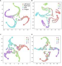
          The experimental results also demonstrated that the proposed type token, sliding window, and local and global multi-head self-attention mechanisms can significantly improve the model's ability to construct, learn, and adaptively select multi-scale spatiotemporal features in MI-EEG signals, and accurately identify electroencephalogram signals in the unilateral motor area. This work provided a novel and accurate classification model for MI-EEG brain-computer interface tasks and proposed a feasible neural rehabilitation assessment scheme based on the model, which could promote the further development and application of MI-EEG methods in neural rehabilitation.

          Related collections

          Most cited references56

          • Record: found
          • Abstract: found
          • Article: not found

          PhysioBank, PhysioToolkit, and PhysioNet

          Circulation, 101(23)
            Bookmark
            • Record: found
            • Abstract: found
            • Article: found
            Is Open Access

            Deep learning with convolutional neural networks for EEG decoding and visualization

            Abstract Deep learning with convolutional neural networks (deep ConvNets) has revolutionized computer vision through end‐to‐end learning, that is, learning from the raw data. There is increasing interest in using deep ConvNets for end‐to‐end EEG analysis, but a better understanding of how to design and train ConvNets for end‐to‐end EEG decoding and how to visualize the informative EEG features the ConvNets learn is still needed. Here, we studied deep ConvNets with a range of different architectures, designed for decoding imagined or executed tasks from raw EEG. Our results show that recent advances from the machine learning field, including batch normalization and exponential linear units, together with a cropped training strategy, boosted the deep ConvNets decoding performance, reaching at least as good performance as the widely used filter bank common spatial patterns (FBCSP) algorithm (mean decoding accuracies 82.1% FBCSP, 84.0% deep ConvNets). While FBCSP is designed to use spectral power modulations, the features used by ConvNets are not fixed a priori. Our novel methods for visualizing the learned features demonstrated that ConvNets indeed learned to use spectral power modulations in the alpha, beta, and high gamma frequencies, and proved useful for spatially mapping the learned features by revealing the topography of the causal contributions of features in different frequency bands to the decoding decision. Our study thus shows how to design and train ConvNets to decode task‐related information from the raw EEG without handcrafted features and highlights the potential of deep ConvNets combined with advanced visualization techniques for EEG‐based brain mapping. Hum Brain Mapp 38:5391–5420, 2017. © 2017 Wiley Periodicals, Inc.
              Bookmark
              • Record: found
              • Abstract: found
              • Article: not found

              EEGNet: a compact convolutional neural network for EEG-based brain–computer interfaces

              Brain-computer interfaces (BCI) enable direct communication with a computer, using neural activity as the control signal. This neural signal is generally chosen from a variety of well-studied electroencephalogram (EEG) signals. For a given BCI paradigm, feature extractors and classifiers are tailored to the distinct characteristics of its expected EEG control signal, limiting its application to that specific signal. Convolutional neural networks (CNNs), which have been used in computer vision and speech recognition to perform automatic feature extraction and classification, have successfully been applied to EEG-based BCIs; however, they have mainly been applied to single BCI paradigms and thus it remains unclear how these architectures generalize to other paradigms. Here, we ask if we can design a single CNN architecture to accurately classify EEG signals from different BCI paradigms, while simultaneously being as compact as possible.
                Bookmark

                Author and article information

                Contributors
                Journal
                Front Neurosci
                Front Neurosci
                Front. Neurosci.
                Frontiers in Neuroscience
                Frontiers Media S.A.
                1662-4548
                1662-453X
                15 August 2023
                2023
                : 17
                : 1204385
                Affiliations
                [1] 1School of Computer Science and Technology, Donghua University , Shanghai, China
                [2] 2Department of Neurosurgery and State Key Laboratory of Trauma, Burn and Combined Injury, Southwest Hospital, Third Military Medical University (Army Medical University) , Chongqing, China
                [3] 3Department of Orthopaedics of TCM Clinical Unit, The Sixth Medical Center, Chinese PLA General Hospital , Beijing, China
                Author notes

                Edited by: Shugeng Chen, Huashan Hospital, Fudan University, China

                Reviewed by: Giulia Cisotto, University of Milano-Bicocca, Italy; Yuhu Shi, Shanghai Maritime University, China

                *Correspondence: Zijian Wang wang.zijian@ 123456dhu.edu.cn
                Article
                10.3389/fnins.2023.1204385
                10469504
                37662108
                72d9304c-8265-4ff4-957f-3962279468f3
                Copyright © 2023 Huang, Zheng, Xu, Li, Liu, Wang, Feng and Cao.

                This is an open-access article distributed under the terms of the Creative Commons Attribution License (CC BY). The use, distribution or reproduction in other forums is permitted, provided the original author(s) and the copyright owner(s) are credited and that the original publication in this journal is cited, in accordance with accepted academic practice. No use, distribution or reproduction is permitted which does not comply with these terms.

                History
                : 12 April 2023
                : 26 July 2023
                Page count
                Figures: 10, Tables: 7, Equations: 12, References: 56, Pages: 17, Words: 10283
                Funding
                This study was supported by the Shanghai Sailing Program (No. 23YF1401100) and the Fundamental Research Funds for the Central Universities (No. 2232021D-26).
                Categories
                Neuroscience
                Original Research
                Custom metadata
                Neural Technology

                Neurosciences
                eeg,motor imagery,brain computer interface,deep learning,cnn,attention
                Neurosciences
                eeg, motor imagery, brain computer interface, deep learning, cnn, attention

                Comments

                Comment on this article