107
views
0
recommends
+1 Recommend
0 collections
    0
    shares
      • Record: found
      • Abstract: found
      • Article: found
      Is Open Access

      Macrophages in inflammatory multiple sclerosis lesions have an intermediate activation status

      research-article

      Read this article at

      Bookmark
          There is no author summary for this article yet. Authors can add summaries to their articles on ScienceOpen to make them more accessible to a non-specialist audience.

          Abstract

          Background

          Macrophages play a dual role in multiple sclerosis (MS) pathology. They can exert neuroprotective and growth promoting effects but also contribute to tissue damage by production of inflammatory mediators. The effector function of macrophages is determined by the way they are activated. Stimulation of monocyte-derived macrophages in vitro with interferon-γ and lipopolysaccharide results in classically activated (CA/M1) macrophages, and activation with interleukin 4 induces alternatively activated (AA/M2) macrophages.

          Methods

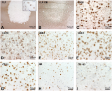
          For this study, the expression of a panel of typical M1 and M2 markers on human monocyte derived M1 and M2 macrophages was analyzed using flow cytometry. This revealed that CD40 and mannose receptor (MR) were the most distinctive markers for human M1 and M2 macrophages, respectively. Using a panel of M1 and M2 markers we next examined the activation status of macrophages/microglia in MS lesions, normal appearing white matter and healthy control samples.

          Results

          Our data show that M1 markers, including CD40, CD86, CD64 and CD32 were abundantly expressed by microglia in normal appearing white matter and by activated microglia and macrophages throughout active demyelinating MS lesions. M2 markers, such as MR and CD163 were expressed by myelin-laden macrophages in active lesions and perivascular macrophages. Double staining with anti-CD40 and anti-MR revealed that approximately 70% of the CD40-positive macrophages in MS lesions also expressed MR, indicating that the majority of infiltrating macrophages and activated microglial cells display an intermediate activation status.

          Conclusions

          Our findings show that, although macrophages in active MS lesions predominantly display M1 characteristics, a major subset of macrophages have an intermediate activation status.

          Related collections

          Most cited references46

          • Record: found
          • Abstract: found
          • Article: not found

          Macrophage activation switching: an asset for the resolution of inflammation.

          Macrophages play a central role in inflammation and host defence against microorganisms, but they also participate actively in the resolution of inflammation after alternative activation. However, it is not known whether the resolution of inflammation requires alternative activation of new resting monocytes/macrophages or if proinflammatory activated macrophages have the capacity to switch their activation towards anti-inflammation. In order to answer this question, we first characterized differential human macrophage activation phenotypes. We found that CD163 and CD206 exhibited mutually exclusive induction patterns after stimulation by a panel of anti-inflammatory molecules, whereas CCL18 showed a third, overlapping, pattern. Hence, alternative activation is not a single process, but provides a variety of different cell populations. The capacity of macrophages to switch from one activation state to another was then assessed by determining the reversibility of CD163 and CD206 expression and of CCL18 and CCL3 production. We found that every activation state was rapidly and fully reversible, suggesting that a given cell may participate sequentially in both the induction and the resolution of inflammation. These findings may provide new insight into the inflammatory process as well as new fields of investigation for immunotherapy in the fields of chronic inflammatory diseases and cancer.
            Bookmark
            • Record: found
            • Abstract: found
            • Article: not found

            Experimental autoimmune encephalomyelitis repressed by microglial paralysis.

            Although microglial activation occurs in inflammatory, degenerative and neoplastic central nervous system (CNS) disorders, its role in pathogenesis is unclear. We studied this question by generating CD11b-HSVTK transgenic mice, which express herpes simplex thymidine kinase in macrophages and microglia. Ganciclovir treatment of organotypic brain slice cultures derived from CD11b-HSVTK mice abolished microglial release of nitrite, proinflammatory cytokines and chemokines. Systemic ganciclovir administration to CD11b-HSVTK mice elicited hematopoietic toxicity, which was prevented by transfer of wild-type bone marrow. In bone marrow chimeras, ganciclovir blocked microglial activation in the facial nucleus upon axotomy and repressed the development of experimental autoimmune encephalomyelitis. We conclude that microglial paralysis inhibits the development and maintenance of inflammatory CNS lesions. The microglial compartment thus provides a potential therapeutic target in inflammatory CNS disorders. These results validate CD11b-HSVTK mice as a tool to study the impact of microglial activation on CNS diseases in vivo.
              Bookmark
              • Record: found
              • Abstract: found
              • Article: not found

              The mononuclear phagocyte system: a new classification of macrophages, monocytes, and their precursor cells.

              There have been many attempts in the past to classify phagocytic mononuclear cells and to define the cell system they are considered to form-among these being the "macrophage system" of Metchnikoff, the "reticulo-endothelial system" of Aschoff, and the "reticulo-histiocyte system" proposed by Volterra and reintroduced by Thomas. None of these is entirely adequate in the light of present knowledge. In 1969, therefore, a group of workers proposed a new classification of all highly phagocytic mononuclear cells and their precursors in what they termed the "mononuclear phagocyte system". This system includes the promonocytes and their precursors in the bone marrow, the monocytes in the peripheral blood, and the macrophages in the tissues. Subsequent consultation with numerous other specialists throughout the world led to a certain number of changes in this classification, which is now proposed in revised form.Inclusion of cells in the "mononuclear phagocyte system" is based on similarities in the morphology, function, origin, and kinetics of the phagocytes. By these criteria reticular cells, dendritic cells, endothelial cells, and fibroblasts (fibrocytes) are excluded. The proponents point out that as new knowledge is acquired modifications may have to be made, certain cells being added to or removed from the new classification.
                Bookmark

                Author and article information

                Journal
                J Neuroinflammation
                J Neuroinflammation
                Journal of Neuroinflammation
                BioMed Central
                1742-2094
                2013
                4 March 2013
                : 10
                : 35
                Affiliations
                [1 ]Department of Molecular Cell Biology and Immunology, VU University Medical Center, Van der Boechhorststraat 7, BT Amsterdam, 1081, The Netherlands
                [2 ]Department of Pathology, VU University Medical Center, De Boelelaan 1117, HV Amsterdam, 1081, The Netherlands
                [3 ]Department of Neuroimmunlogy Unit, Blizard Institute, Barts and the London School of Medicine Dentistry, Queen Mary University of London, 4 Newark Street, London, E1 2AT, United Kingdom
                [4 ]Department of Clinical Chemistry, VU University Medical Center, De Boelelaan 1117, HV Amsterdam, 1081, The Netherlands
                Article
                1742-2094-10-35
                10.1186/1742-2094-10-35
                3610294
                23452918
                78004840-2e4e-4a97-a4a5-74a61fe98a31
                Copyright ©2013 Vogel et al.; licensee BioMed Central Ltd.

                This is an Open Access article distributed under the terms of the Creative Commons Attribution License ( http://creativecommons.org/licenses/by/2.0), which permits unrestricted use, distribution, and reproduction in any medium, provided the original work is properly cited.

                History
                : 21 January 2013
                : 22 February 2013
                Categories
                Research

                Neurosciences
                cd40,macrophages,mannose receptor,multiple sclerosis
                Neurosciences
                cd40, macrophages, mannose receptor, multiple sclerosis

                Comments

                Comment on this article