Inviting an author to review:
Find an author and click ‘Invite to review selected article’ near their name.
Search for authorsSearch for similar articles
25
views
0
recommends
+1 Recommend
0 collections
    0
    shares
      • Record: found
      • Abstract: found
      • Article: found
      Is Open Access

      Heparan sulfate proteoglycans (HSPGs) and chondroitin sulfate proteoglycans (CSPGs) function as endocytic receptors for an internalizing anti-nucleic acid antibody

      research-article

      Read this article at

      Bookmark
          There is no author summary for this article yet. Authors can add summaries to their articles on ScienceOpen to make them more accessible to a non-specialist audience.

          Abstract

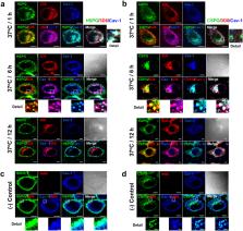
          A subset of monoclonal anti-DNA autoantibodies enters a variety of living cells. Here, we aimed to identify the endocytic receptors recognized by an internalizing anti-nucleic acid autoantibody, the 3D8 single-chain variable fragment (scFv). We found that cell surface binding and internalization of 3D8 scFv were inhibited markedly in soluble heparan sulfate (HS)/chondroitin sulfate (CS)-deficient or -removed cells and in the presence of soluble HS and CS. 3D8 scFv colocalized intracellularly with either HS proteoglycans (HSPGs) or CSPGs in HeLa cells. 3D8 scFv was co-endocytosed and co-precipitated with representative individual HSPG and CSPG molecules: syndecan-2 (a transmembrane HSPG), glypican-3 (a glycosylphosphatidylinositol (GPI)-anchored HSPG); CD44 (a transmembrane CSPG); and brevican (a GPI-anchored CSPG). Collected data indicate that 3D8 scFv binds to the negatively charged sugar chains of both HSPGs and CSPGs and is then internalized along with these molecules, irrespective of how these proteoglycans are associated with the cell membrane. This is the first study to show that anti-DNA antibodies enter cells via both HSPGs and CSPGs simultaneously. The data may aid understanding of endocytic receptors that bind anti-DNA autoantibodies. The study also provides insight into potential cell membrane targets for macromolecular delivery.

          Related collections

          Most cited references42

          • Record: found
          • Abstract: found
          • Article: not found

          Cancer cell exosomes depend on cell-surface heparan sulfate proteoglycans for their internalization and functional activity.

          Extracellular vesicle (EV)-mediated intercellular transfer of signaling proteins and nucleic acids has recently been implicated in the development of cancer and other pathological conditions; however, the mechanism of EV uptake and how this may be targeted remain as important questions. Here, we provide evidence that heparan sulfate (HS) proteoglycans (PGs; HSPGs) function as internalizing receptors of cancer cell-derived EVs with exosome-like characteristics. Internalized exosomes colocalized with cell-surface HSPGs of the syndecan and glypican type, and exosome uptake was specifically inhibited by free HS chains, whereas closely related chondroitin sulfate had no effect. By using several cell mutants, we provide genetic evidence of a receptor function of HSPG in exosome uptake, which was dependent on intact HS, specifically on the 2-O and N-sulfation groups. Further, enzymatic depletion of cell-surface HSPG or pharmacological inhibition of endogenous PG biosynthesis by xyloside significantly attenuated exosome uptake. We provide biochemical evidence that HSPGs are sorted to and associate with exosomes; however, exosome-associated HSPGs appear to have no direct role in exosome internalization. On a functional level, exosome-induced ERK1/2 signaling activation was attenuated in PG-deficient mutant cells as well as in WT cells treated with xyloside. Importantly, exosome-mediated stimulation of cancer cell migration was significantly reduced in PG-deficient mutant cells, or by treatment of WT cells with heparin or xyloside. We conclude that cancer cell-derived exosomes use HSPGs for their internalization and functional activity, which significantly extends the emerging role of HSPGs as key receptors of macromolecular cargo.
            Bookmark
            • Record: found
            • Abstract: found
            • Article: not found

            Heparan sulfate proteoglycans mediate internalization and propagation of specific proteopathic seeds.

            Recent experimental evidence suggests that transcellular propagation of fibrillar protein aggregates drives the progression of neurodegenerative diseases in a prion-like manner. This phenomenon is now well described in cell and animal models and involves the release of protein aggregates into the extracellular space. Free aggregates then enter neighboring cells to seed further fibrillization. The mechanism by which aggregated extracellular proteins such as tau and α-synuclein bind and enter cells to trigger intracellular fibril formation is unknown. Prior work indicates that prion protein aggregates bind heparan sulfate proteoglycans (HSPGs) on the cell surface to transmit pathologic processes. Here, we find that tau fibril uptake also occurs via HSPG binding. This is blocked in cultured cells and primary neurons by heparin, chlorate, heparinase, and genetic knockdown of a key HSPG synthetic enzyme, Ext1. Interference with tau binding to HSPGs prevents recombinant tau fibrils from inducing intracellular aggregation and blocks transcellular aggregate propagation. In vivo, a heparin mimetic, F6, blocks neuronal uptake of stereotactically injected tau fibrils. Finally, uptake and seeding by α-synuclein fibrils, but not huntingtin fibrils, occurs by the same mechanism as tau. This work suggests a unifying mechanism of cell uptake and propagation for tauopathy and synucleinopathy.
              Bookmark
              • Record: found
              • Abstract: found
              • Article: not found

              Heparin and heparan sulfate: structure and function.

              This review covers the structure and function of heparin and heparan sulfate glycosaminoglycans. Their chemical structures are discussed, including recently developed methods for sequencing picomole to nanomole quantities of heparin- and heparan sulfate-derived oligosaccharides. The biosynthesis of heparin and heparan sulfate is reviewed as it relates to their diverse and varied structures, and their biological activities and functions are discussed. The literature up to August 2001 is reviewed, and 208 references are cited.
                Bookmark

                Author and article information

                Contributors
                kwonmh@ajou.ac.kr
                Journal
                Sci Rep
                Sci Rep
                Scientific Reports
                Nature Publishing Group UK (London )
                2045-2322
                30 October 2017
                30 October 2017
                2017
                : 7
                : 14373
                Affiliations
                [1 ]ISNI 0000 0004 0532 3933, GRID grid.251916.8, Department of Biomedical Sciences, , Graduate School, Ajou University, ; 206 World cup-ro, Yeongtong-gu, Suwon, 443-749 South Korea
                [2 ]ISNI 0000 0004 0532 3933, GRID grid.251916.8, Department of Microbiology, , Ajou University School of Medicine, ; 206 World cup-ro, Yeongtong-gu, Suwon, 443-749 South Korea
                [3 ]Animal Biotechnology Division, National Institute of Animal Science, RDA, 1500, Kongjwipatjwi-ro, Iseo-myeon, Wanju-gun, Jeollabuk-do 565-851 South Korea
                Author information
                http://orcid.org/0000-0001-8757-3693
                Article
                14793
                10.1038/s41598-017-14793-z
                5662561
                29085061
                7cb391f7-e8bf-40c5-a12d-7ea23dd5b961
                © The Author(s) 2017

                Open Access This article is licensed under a Creative Commons Attribution 4.0 International License, which permits use, sharing, adaptation, distribution and reproduction in any medium or format, as long as you give appropriate credit to the original author(s) and the source, provide a link to the Creative Commons license, and indicate if changes were made. The images or other third party material in this article are included in the article’s Creative Commons license, unless indicated otherwise in a credit line to the material. If material is not included in the article’s Creative Commons license and your intended use is not permitted by statutory regulation or exceeds the permitted use, you will need to obtain permission directly from the copyright holder. To view a copy of this license, visit http://creativecommons.org/licenses/by/4.0/.

                History
                : 3 March 2017
                : 16 October 2017
                Categories
                Article
                Custom metadata
                © The Author(s) 2017

                Uncategorized
                Uncategorized

                Comments

                Comment on this article