22
views
0
recommends
+1 Recommend
0 collections
    0
    shares
      • Record: found
      • Abstract: found
      • Article: found
      Is Open Access

      Organ-Protective Effects and the Underlying Mechanism of Dexmedetomidine

      review-article
      ,
      Mediators of Inflammation
      Hindawi

      Read this article at

      Bookmark
          There is no author summary for this article yet. Authors can add summaries to their articles on ScienceOpen to make them more accessible to a non-specialist audience.

          Abstract

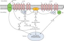
          Dexmedetomidine (DEX) is a highly selective α2 adrenergic receptor ( α2AR) agonist currently used in clinical settings. Because DEX has dose-dependent advantages of sedation, analgesia, antianxiety, inhibition of sympathetic nervous system activity, cardiovascular stabilization, and significant reduction of postoperative delirium and agitation, but does not produce respiratory depression and agitation, it is widely used in clinical anesthesia and ICU departments. In recent years, much clinical study and basic research has confirmed that DEX has a protective effect on a variety of organs, including the nervous system, heart, lungs, kidneys, liver, and small intestine. It acts by reducing the inflammatory response in these organs, activating antiapoptotic signaling pathways which protect cells from damage. Therefore, based on wide clinical application and safety, DEX may become a promising clinical multiorgan protection drug in the future. In this article, we review the physiological effects related to organ protection in α2AR agonists along with the organ-protective effects and mechanisms of DEX to understand their combined application value.

          Related collections

          Most cited references97

          • Record: found
          • Abstract: found
          • Article: not found

          NF-kappaB in renal inflammation.

          The NF-kappaB family of transcription factors regulates the induction and resolution of inflammation. Two main pathways, classical and alternative, control the nuclear translocation of NF-kappaB. Classical NF-kappaB activation is usually a rapid and transient response to a wide range of stimuli whose main effector is RelA/p50. The alternative NF-kappaB pathway is a more delayed response to a smaller range of stimuli resulting in DNA binding of RelB/p52 complexes. Additional complexity in this system involves the posttranslational modification of NF-kappaB proteins and an ever-increasing range of co-activators, co-repressors, and NF-kappaB complex proteins. Collectively, NF-kappaB regulates the expression of numerous genes that play a key role in the inflammatory response during human and experimental kidney injury. Multiple stimuli activate NF-kappaB through the classical pathway in somatic renal cells, and noncanonical pathway activation by TWEAK occurs in acute kidney injury. Under most test conditions, specific NF-kappaB inhibitors tend to reduce inflammation in experimental kidney injury but not always. Although many drugs in current use clinically influence NF-kappaB activation, there are no data regarding specific NF-kappaB inhibition in human kidney disease.
            Bookmark
            • Record: found
            • Abstract: found
            • Article: not found

            Oxygen free radicals in ischemic acute renal failure in the rat.

            During renal ischemia, ATP is degraded to hypoxanthine. When xanthine oxidase converts hypoxanthine to xanthine in the presence of molecular oxygen, superoxide radical (O-2) is generated. We studied the role of O-2 and its reduction product OH X in mediating renal injury after ischemia. Male Sprague-Dawley rats underwent right nephrectomy followed by 60 min of occlusion of the left renal artery. The O-2 scavenger superoxide dismutase (SOD) was given 8 min before clamping and before release of the renal artery clamp. Control rats received 5% dextrose instead. Plasma creatinine was lower in SOD treated rats: 1.5, 1.0, and 0.8 mg/dl vs. 2.5, 2.5, and 2.1 mg/dl at 24, 48, and 72 h postischemia. 24 h after ischemia inulin clearance was higher in SOD treated rats than in controls (399 vs. 185 microliter/min). Renal blood flow, measured after ischemia plus 15 min of reflow, was also greater in SOD treated than in control rats. Furthermore, tubular injury, judged histologically in perfusion fixed specimens, was less in SOD treated rats. Rats given SOD inactivated by prior incubation with diethyldithiocarbamate had plasma creatinine values no different from those of control rats. The OH X scavenger dimethylthiourea (DMTU) was given before renal artery occlusion. DMTU treated rats had lower plasma creatinine than did controls: 1.7, 1.7, and 1.3 mg/dl vs. 3.2, 2.2, and 2.4 mg/dl at 24, 48, and 72 h postischemia. Neither SOD nor DMTU caused an increase in renal blood flow, urine flow rate, or solute excretion in normal rats. The xanthine oxidase inhibitor allopurinol was given before ischemia to prevent the generation of oxygen free radicals. Plasma creatinine was lower in allopurinol treated rats: 2.7, 2.2, and 1.4 mg/dl vs. 3.6, 3.5, and 2.3 mg/dl at 24, 48, and 72 h postischemia. Catalase treatment did not protect against renal ischemia, perhaps because its large size limits glomerular filtration and access to the tubular lumen. Superoxide-mediated lipid peroxidation was studied after renal ischemia. 60 min of ischemia did not increase the renal content of the lipid peroxide malondialdehyde, whereas ischemia plus 15 min reflow resulted in a large increase in kidney lipid peroxides. Treatment with SOD before renal ischemia prevented the reflow-induced increase in lipid peroxidation in renal cortical mitochondria but not in crude cortical homogenates. In summary, the oxygen free radical scavengers SOD and DMTU, and allopurinol, which inhibits free radical generation, protected renal function after ischemia. Reperfusion after ischemia resulted in lipid peroxidation; SOD decreased lipid peroxidation in cortical mitochondria after renal ischemia and reflow. We concluded that restoration of oxygen supply to ischemic kidney results in the production of oxygen free radicals, which causes renal injury by lipid peroxidation.
              Bookmark
              • Record: found
              • Abstract: found
              • Article: not found

              Diversity of G proteins in signal transduction

              The heterotrimeric guanine nucleotide-binding proteins (G proteins) act as switches that regulate information processing circuits connecting cell surface receptors to a variety of effectors. The G proteins are present in all eukaryotic cells, and they control metabolic, humoral, neural, and developmental functions. More than a hundred different kinds of receptors and many different effectors have been described. The G proteins that coordinate receptor-effector activity are derived from a large gene family. At present, the family is known to contain at least sixteen different genes that encode the alpha subunit of the heterotrimer, four that encode beta subunits, and multiple genes encoding gamma subunits. Specific transient interactions between these components generate the pathways that modulate cellular responses to complex chemical signals.
                Bookmark

                Author and article information

                Contributors
                Journal
                Mediators Inflamm
                Mediators Inflamm
                MI
                Mediators of Inflammation
                Hindawi
                0962-9351
                1466-1861
                2020
                9 May 2020
                : 2020
                : 6136105
                Affiliations
                Department of Anesthesiology, The First Hospital of China Medical University, 155 Nanjing North Street, Shenyang, China
                Author notes

                Guest Editor: Xiaolu Jin

                Author information
                https://orcid.org/0000-0001-7120-4789
                https://orcid.org/0000-0002-1162-9686
                Article
                10.1155/2020/6136105
                7232715
                32454792
                8089673f-96bb-4c49-8e90-beb4631ad515
                Copyright © 2020 Naren Bao and Bing Tang.

                This is an open access article distributed under the Creative Commons Attribution License, which permits unrestricted use, distribution, and reproduction in any medium, provided the original work is properly cited.

                History
                : 3 March 2020
                : 13 April 2020
                : 16 April 2020
                Funding
                Funded by: Department of Education of Liaoning Province
                Award ID: LK201651
                Categories
                Review Article

                Immunology
                Immunology

                Comments

                Comment on this article