1
views
0
recommends
+1 Recommend
0 collections
    0
    shares
      • Record: found
      • Abstract: found
      • Article: found
      Is Open Access

      Communication Between Cardiomyocytes and Fibroblasts During Cardiac Ischemia/Reperfusion and Remodeling: Roles of TGF-β, CTGF, the Renin Angiotensin Axis, and Non-coding RNA Molecules

      review-article

      Read this article at

      Bookmark
          There is no author summary for this article yet. Authors can add summaries to their articles on ScienceOpen to make them more accessible to a non-specialist audience.

          Abstract

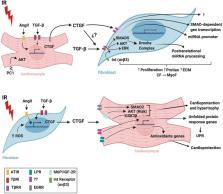
          Communication between cells is a foundational concept for understanding the physiology and pathology of biological systems. Paracrine/autocrine signaling, direct cell-to-cell interplay, and extracellular matrix interactions are three types of cell communication that regulate responses to different stimuli. In the heart, cardiomyocytes, fibroblasts, and endothelial cells interact to form the cardiac tissue. Under pathological conditions, such as myocardial infarction, humoral factors released by these cells may induce tissue damage or protection, depending on the type and concentration of molecules secreted. Cardiac remodeling is also mediated by the factors secreted by cardiomyocytes and fibroblasts that are involved in the extensive reciprocal interactions between these cells. Identifying the molecules and cellular signal pathways implicated in these processes will be crucial for creating effective tissue-preserving treatments during or after reperfusion. Numerous therapies to protect cardiac tissue from reperfusion-induced injury have been explored, and ample pre-clinical research has attempted to identify drugs or techniques to mitigate cardiac damage. However, despite great success in animal models, it has not been possible to completely translate these cardioprotective effects to human applications. This review provides a current summary of the principal molecules, pathways, and mechanisms underlying cardiomyocyte and cardiac fibroblast crosstalk during ischemia/reperfusion injury. We also discuss pre-clinical molecules proposed as treatments for myocardial infarction and provide a clinical perspective on these potential therapeutic agents.

          Related collections

          Most cited references168

          • Record: found
          • Abstract: found
          • Article: not found

          Dysregulation of microRNAs after myocardial infarction reveals a role of miR-29 in cardiac fibrosis.

          Acute myocardial infarction (MI) due to coronary artery occlusion is accompanied by a pathological remodeling response that includes hypertrophic cardiac growth and fibrosis, which impair cardiac contractility. Previously, we showed that cardiac hypertrophy and heart failure are accompanied by characteristic changes in the expression of a collection of specific microRNAs (miRNAs), which act as negative regulators of gene expression. Here, we show that MI in mice and humans also results in the dysregulation of specific miRNAs, which are similar to but distinct from those involved in hypertrophy and heart failure. Among the MI-regulated miRNAs are members of the miR-29 family, which are down-regulated in the region of the heart adjacent to the infarct. The miR-29 family targets a cadre of mRNAs that encode proteins involved in fibrosis, including multiple collagens, fibrillins, and elastin. Thus, down-regulation of miR-29 would be predicted to derepress the expression of these mRNAs and enhance the fibrotic response. Indeed, down-regulation of miR-29 with anti-miRs in vitro and in vivo induces the expression of collagens, whereas over-expression of miR-29 in fibroblasts reduces collagen expression. We conclude that miR-29 acts as a regulator of cardiac fibrosis and represents a potential therapeutic target for tissue fibrosis in general.
            Bookmark
            • Record: found
            • Abstract: found
            • Article: not found

            The Biological Basis for Cardiac Repair After Myocardial Infarction: From Inflammation to Fibrosis.

            In adult mammals, massive sudden loss of cardiomyocytes after infarction overwhelms the limited regenerative capacity of the myocardium, resulting in the formation of a collagen-based scar. Necrotic cells release danger signals, activating innate immune pathways and triggering an intense inflammatory response. Stimulation of toll-like receptor signaling and complement activation induces expression of proinflammatory cytokines (such as interleukin-1 and tumor necrosis factor-α) and chemokines (such as monocyte chemoattractant protein-1/ chemokine (C-C motif) ligand 2 [CCL2]). Inflammatory signals promote adhesive interactions between leukocytes and endothelial cells, leading to extravasation of neutrophils and monocytes. As infiltrating leukocytes clear the infarct from dead cells, mediators repressing inflammation are released, and anti-inflammatory mononuclear cell subsets predominate. Suppression of the inflammatory response is associated with activation of reparative cells. Fibroblasts proliferate, undergo myofibroblast transdifferentiation, and deposit large amounts of extracellular matrix proteins maintaining the structural integrity of the infarcted ventricle. The renin-angiotensin-aldosterone system and members of the transforming growth factor-β family play an important role in activation of infarct myofibroblasts. Maturation of the scar follows, as a network of cross-linked collagenous matrix is formed and granulation tissue cells become apoptotic. This review discusses the cellular effectors and molecular signals regulating the inflammatory and reparative response after myocardial infarction. Dysregulation of immune pathways, impaired suppression of postinfarction inflammation, perturbed spatial containment of the inflammatory response, and overactive fibrosis may cause adverse remodeling in patients with infarction contributing to the pathogenesis of heart failure. Therapeutic modulation of the inflammatory and reparative response may hold promise for the prevention of postinfarction heart failure.
              Bookmark
              • Record: found
              • Abstract: found
              • Article: not found

              Acute myocardial infarction.

              Acute myocardial infarction has traditionally been divided into ST elevation or non-ST elevation myocardial infarction; however, therapies are similar between the two, and the overall management of acute myocardial infarction can be reviewed for simplicity. Acute myocardial infarction remains a leading cause of morbidity and mortality worldwide, despite substantial improvements in prognosis over the past decade. The progress is a result of several major trends, including improvements in risk stratification, more widespread use of an invasive strategy, implementation of care delivery systems prioritising immediate revascularisation through percutaneous coronary intervention (or fibrinolysis), advances in antiplatelet agents and anticoagulants, and greater use of secondary prevention strategies such as statins. This seminar discusses the important topics of the pathophysiology, epidemiological trends, and modern management of acute myocardial infarction, focusing on the recent advances in reperfusion strategies and pharmacological treatment approaches.
                Bookmark

                Author and article information

                Contributors
                Journal
                Front Physiol
                Front Physiol
                Front. Physiol.
                Frontiers in Physiology
                Frontiers Media S.A.
                1664-042X
                03 September 2021
                2021
                : 12
                : 716721
                Affiliations
                [1] 1Advanced Center for Chronic Diseases (ACCDiS), Facultad de Ciencias Químicas y Farmacéuticas & Facultad de Medicina, Universidad de Chile , Santiago de Chile, Chile
                [2] 2Programa de Fisiología y Biofísica, Instituto de Ciencias Biomédicas (ICBM), Facultad de Medicina, Universidad de Chile , Santiago de Chile, Chile
                [3] 3Programa de Fisiopatología, Instituto de Ciencias Biomédicas (ICBM), Facultad de Medicina, Universidad de Chile , Santiago de Chile, Chile
                [4] 4Red para el Estudio de Enfermedades Cardiopulmonares de alta letalidad (REECPAL), Universidad de Chile , Santiago de Chile, Chile
                [5] 5Escuela de Química y Farmacia, Facultad de Medicina, Universidad Andrés Bello , Viña del Mar, Chile
                [6] 6Departamento de Química Farmacológica y Toxicológica, Facultad de Ciencias Químicas y Farmacéuticas, Universidad de Chile , Santiago de Chile, Chile
                [7] 7Programa de Farmacología Molecular y Clínica, Instituto de Ciencias Biomédicas, Facultad de Medicina, Universidad de Chile , Santiago de Chile, Chile
                Author notes

                Edited by: Rodrigo Andres Carrasco Loza, Peter Munk Cardiac Centre, Canada

                Reviewed by: Moni Nader, Khalifa University, United Arab Emirates; Esma Nur Okatan, University of Istinye, Turkey

                *Correspondence: Zully Pedrozo, zpedrozo@ 123456uchile.cl

                These authors have contributed equally to this work

                This article was submitted to Integrative Physiology, a section of the journal Frontiers in Physiology

                Article
                10.3389/fphys.2021.716721
                8446518
                34539441
                84eb697d-1acb-41d8-8de4-bc1995af9a7c
                Copyright © 2021 Flores-Vergara, Olmedo, Aránguiz, Riquelme, Vivar and Pedrozo.

                This is an open-access article distributed under the terms of the Creative Commons Attribution License (CC BY). The use, distribution or reproduction in other forums is permitted, provided the original author(s) and the copyright owner(s) are credited and that the original publication in this journal is cited, in accordance with accepted academic practice. No use, distribution or reproduction is permitted which does not comply with these terms.

                History
                : 29 May 2021
                : 26 July 2021
                Page count
                Figures: 4, Tables: 0, Equations: 0, References: 168, Pages: 16, Words: 13605
                Funding
                Funded by: Fondo Nacional de Desarrollo Científico y Tecnólogico
                Award ID: FONDECYT 1180613
                Award ID: 11181000
                Award ID: REDES 170032
                Award ID: FONDAP 15130011
                Funded by: Universidad Andrés Bello 10.13039/501100013908
                Award ID: DI-02-20/REG
                Categories
                Physiology
                Review

                Anatomy & Physiology
                myocardial infarction,ischemia/reperfusion,crosstalk,fibroblasts,cardiomyocytes

                Comments

                Comment on this article