20
views
0
recommends
+1 Recommend
0 collections
    0
    shares
      • Record: found
      • Abstract: found
      • Article: found
      Is Open Access

      Distinct Dorsal and Ventral Hippocampal CA3 Outputs Govern Contextual Fear Discrimination

      research-article
      1 , 2 , 3 , 1 , 2 , 3 , 1 , 2 , 3 , 4 , 5 , *
      Cell reports

      Read this article at

      Bookmark
          There is no author summary for this article yet. Authors can add summaries to their articles on ScienceOpen to make them more accessible to a non-specialist audience.

          SUMMARY

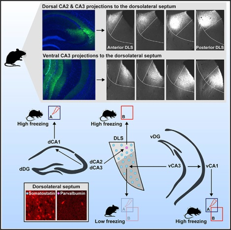
          Considerable work emphasizes a role for hippocampal circuits in governing contextual fear discrimination. However· the intra- and extrahippocampal pathways that route contextual information to cortical and subcortical circuits to guide adaptive behavioral responses are poorly understood. Using terminal-specific optogenetic silencing in a contextual fear discrimination learning paradigm· we identify opposing roles for dorsal CA3-CA1 (dCA3-dCA1) projections and dorsal CA3-dorsolateral septum (dCA3-DLS) projections in calibrating fear responses to certain and ambiguous contextual threats· respectively. Ventral CA3-DLS (vCA3-DLS) projections suppress fear responses in both certain and ambiguous contexts· whereas ventral CA3-CA1 (vCA3-vCA1) projections promote fear responses in both these contexts. Lastly· using retrograde monosynaptic tracing· ex vivo electrophysiological recordings· and optogenetics,· we identify a sparse population of DLS parvalbumin (PV) neurons as putative relays of dCA3-DLS projections to diverse subcortical circuits. Taken together· these studies illuminate how distinct dCA3 and vCA3 outputs calibrate contextual fear discrimination.

          Graphical Abstract

          In Brief

          Besnard et al. show that dorsal and ventral hippocampal CA3 projections to CA1 and dorsolateral septum (DLS) play distinct roles in calibration of contextual fear discrimination. DLS parvalbumin inhibitory neurons receive monosynaptic dorsal CA3 inputs and modulate fear responses in a context-specific manner.

          Related collections

          Most cited references54

          • Record: found
          • Abstract: found
          • Article: not found

          A resource of Cre driver lines for genetic targeting of GABAergic neurons in cerebral cortex.

          A key obstacle to understanding neural circuits in the cerebral cortex is that of unraveling the diversity of GABAergic interneurons. This diversity poses general questions for neural circuit analysis: how are these interneuron cell types generated and assembled into stereotyped local circuits and how do they differentially contribute to circuit operations that underlie cortical functions ranging from perception to cognition? Using genetic engineering in mice, we have generated and characterized approximately 20 Cre and inducible CreER knockin driver lines that reliably target major classes and lineages of GABAergic neurons. More select populations are captured by intersection of Cre and Flp drivers. Genetic targeting allows reliable identification, monitoring, and manipulation of cortical GABAergic neurons, thereby enabling a systematic and comprehensive analysis from cell fate specification, migration, and connectivity, to their functions in network dynamics and behavior. As such, this approach will accelerate the study of GABAergic circuits throughout the mammalian brain. Copyright © 2011 Elsevier Inc. All rights reserved.
            Bookmark
            • Record: found
            • Abstract: found
            • Article: not found

            Pattern separation in the dentate gyrus and CA3 of the hippocampus.

            Theoretical models have long pointed to the dentate gyrus as a possible source of neuronal pattern separation. In agreement with predictions from these models, we show that minimal changes in the shape of the environment in which rats are exploring can substantially alter correlated activity patterns among place-modulated granule cells in the dentate gyrus. When the environments are made more different, new cell populations are recruited in CA3 but not in the dentate gyrus. These results imply a dual mechanism for pattern separation in which signals from the entorhinal cortex can be decorrelated both by changes in coincidence patterns in the dentate gyrus and by recruitment of nonoverlapping cell assemblies in CA3.
              Bookmark
              • Record: found
              • Abstract: found
              • Article: not found

              Pattern separation in the hippocampus.

              The ability to discriminate among similar experiences is a crucial feature of episodic memory. This ability has long been hypothesized to require the hippocampus, and computational models suggest that it is dependent on pattern separation. However, empirical data for the role of the hippocampus in pattern separation have not been available until recently. This review summarizes data from electrophysiological recordings, lesion studies, immediate-early gene imaging, transgenic mouse models, as well as human functional neuroimaging, that provide convergent evidence for the involvement of particular hippocampal subfields in this key process. We discuss the impact of aging and adult neurogenesis on pattern separation, and also highlight several challenges to linking across species and approaches, and suggest future directions for investigation. Copyright © 2011 Elsevier Ltd. All rights reserved.
                Bookmark

                Author and article information

                Journal
                101573691
                39703
                Cell Rep
                Cell Rep
                Cell reports
                2211-1247
                21 February 2020
                18 February 2020
                02 March 2020
                : 30
                : 7
                : 2360-2373.e5
                Affiliations
                [1 ]Center for Regenerative Medicine, Massachusetts General Hospital, Boston, MA 02114, USA
                [2 ]Harvard Stem Cell Institute, Cambridge, MA 02138, USA
                [3 ]Department of Psychiatry, Massachusetts General Hospital, Harvard Medical School, Boston, MA 02114, USA
                [4 ]BROAD Institute of Harvard and MIT, Cambridge, MA 02142, USA
                [5 ]Lead Contact
                Author notes

                AUTHOR CONTRIBUTIONS

                A.B. and S.M.M. performed experiments. A.S. and A.B. co-developed the concept, analyzed data, and wrote the manuscript. A.S. conceived the project and supervised all aspects of the project.

                [* ] Correspondence: asahay@ 123456mgh.harvard.edu
                Article
                NIHMS1563625
                10.1016/j.celrep.2020.01.055
                7050277
                32075769
                8d097dc1-95fd-4796-97a4-ec989d50cbb3

                This is an open access article under the CC BY-NC-ND license ( http://creativecommons.org/licenses/by-nc-nd/4.0/).

                History
                Categories
                Article

                Cell biology
                Cell biology

                Comments

                Comment on this article