2
views
0
recommends
+1 Recommend
0 collections
    0
    shares
      • Record: found
      • Abstract: found
      • Article: found
      Is Open Access

      The Complexities of Organ Crosstalk in Phosphate Homeostasis: Time to Put Phosphate Sensing Back in the Limelight

      review-article

      Read this article at

      Bookmark
          There is no author summary for this article yet. Authors can add summaries to their articles on ScienceOpen to make them more accessible to a non-specialist audience.

          Abstract

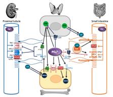
          Phosphate homeostasis is essential for health and is achieved via interaction between the bone, kidney, small intestine, and parathyroid glands and via intricate processes involving phosphate transporters, phosphate sensors, and circulating hormones. Numerous genetic and acquired disorders are associated with disruption in these processes and can lead to significant morbidity and mortality. The role of the kidney in phosphate homeostasis is well known, although it is recognized that the cellular mechanisms in murine models and humans are different. Intestinal phosphate transport also appears to differ in humans and rodents, with recent studies demonstrating a dominant role for the paracellular pathway. The existence of phosphate sensing has been acknowledged for decades; however, the underlying molecular mechanisms are poorly understood. At least three phosphate sensors have emerged. PiT2 and FGFR1c both act as phosphate sensors controlling Fibroblast Growth Factor 23 secretion in bone, whereas the calcium-sensing receptor controls parathyroid hormone secretion in response to extracellular phosphate. All three of the proposed sensors are expressed in the kidney and intestine but their exact function in these organs is unknown. Understanding organ interactions and the mechanisms involved in phosphate sensing requires significant research to develop novel approaches for the treatment of phosphate homeostasis disorders.

          Related collections

          Most cited references89

          • Record: found
          • Abstract: found
          • Article: not found

          The Fibroblast Growth Factor signaling pathway

          The signaling component of the mammalian Fibroblast Growth Factor (FGF) family is comprised of eighteen secreted proteins that interact with four signaling tyrosine kinase FGF receptors (FGFRs). Interaction of FGF ligands with their signaling receptors is regulated by protein or proteoglycan cofactors and by extracellular binding proteins. Activated FGFRs phosphorylate specific tyrosine residues that mediate interaction with cytosolic adaptor proteins and the RAS-MAPK, PI3K-AKT, PLCγ, and STAT intracellular signaling pathways. Four structurally related intracellular non-signaling FGFs interact with and regulate the family of voltage gated sodium channels. Members of the FGF family function in the earliest stages of embryonic development and during organogenesis to maintain progenitor cells and mediate their growth, differentiation, survival, and patterning. FGFs also have roles in adult tissues where they mediate metabolic functions, tissue repair, and regeneration, often by reactivating developmental signaling pathways. Consistent with the presence of FGFs in almost all tissues and organs, aberrant activity of the pathway is associated with developmental defects that disrupt organogenesis, impair the response to injury, and result in metabolic disorders, and cancer. © 2015 Wiley Periodicals, Inc.
            Bookmark
            • Record: found
            • Abstract: found
            • Article: not found

            KDIGO clinical practice guideline for the diagnosis, evaluation, prevention, and treatment of Chronic Kidney Disease-Mineral and Bone Disorder (CKD-MBD).

            (2009)
            The 2009 Kidney Disease: Improving Global Outcomes (KDIGO) clinical practice guideline on the management of chronic kidney disease-mineral and bone disorder (CKD-MBD) is intended to assist the practitioner caring for adults and children with CKD stages 3-5, on chronic dialysis therapy, or with a kidney transplant. The guideline contains recommendations on evaluation and treatment for abnormalities of CKD-MBD. This disease concept of CKD-MBD is based on a prior KDIGO consensus conference. Tests considered are those that relate to the detection and monitoring of laboratory, bone, and cardiovascular abnormalities. Treatments considered are interventions to treat hyperphosphatemia, hyperparathyroidism, and bone disease in patients with CKD stages 3-5D and 1-5T. The guideline development process followed an evidence based approach and treatment recommendations are based on systematic reviews of relevant treatment trials. Recommendations for testing used evidence based on diagnostic accuracy or risk prediction and linked it indirectly with how this would be expected to achieve better outcomes for patients through better detection, evaluation or treatment of disease. Critical appraisal of the quality of the evidence and the strength of recommendations followed the GRADE approach. An ungraded statement was provided when a question did not lend itself to systematic literature review. Limitations of the evidence, especially the lack of definitive clinical outcome trials, are discussed and suggestions are provided for future research.
              Bookmark
              • Record: found
              • Abstract: found
              • Article: not found

              Mechanisms of vascular calcification in chronic kidney disease.

              Vascular calcification is common in chronic kidney disease and associated with increased morbidity and mortality. Its mechanism is multifactorial and incompletely understood. Patients with chronic kidney disease are at risk for vascular calcification because of multiple risk factors that induce vascular smooth muscle cells to change into a chondrocyte or osteoblast-like cell; high total body burden of calcium and phosphorus due to abnormal bone metabolism; low levels of circulating and locally produced inhibitors; impaired renal excretion; and current therapies. Together these factors increase risk and complicate the management of vascular calcification.
                Bookmark

                Author and article information

                Contributors
                Role: Academic Editor
                Journal
                Int J Mol Sci
                Int J Mol Sci
                ijms
                International Journal of Molecular Sciences
                MDPI
                1422-0067
                27 May 2021
                June 2021
                : 22
                : 11
                : 5701
                Affiliations
                [1 ]Department of Neuroscience, Physiology and Pharmacology, Royal Free Campus, University College London, London NW3 2PF, UK; joanne.marks@ 123456ucl.ac.uk
                [2 ]CHU de Nantes, Université de Nantes, F-44042 Nantes, France
                [3 ]Inserm, UMR 1229, RMeS Regenerative Medicine and Skeleton, Université de Nantes, ONIRIS, F-44042 Nantes, France; sarah.beck@ 123456univ-nantes.fr (S.B.-C.); laurent.beck@ 123456inserm.fr (L.B.)
                Author notes
                Author information
                https://orcid.org/0000-0002-6730-2762
                https://orcid.org/0000-0002-2677-2477
                Article
                ijms-22-05701
                10.3390/ijms22115701
                8199323
                34071837
                975f2eb4-489f-41f7-8558-f2032fa271a0
                © 2021 by the authors.

                Licensee MDPI, Basel, Switzerland. This article is an open access article distributed under the terms and conditions of the Creative Commons Attribution (CC BY) license ( https://creativecommons.org/licenses/by/4.0/).

                History
                : 21 March 2021
                : 21 May 2021
                Categories
                Review

                Molecular biology
                phosphate transporter,slc34,slc20,phosphate homeostasis
                Molecular biology
                phosphate transporter, slc34, slc20, phosphate homeostasis

                Comments

                Comment on this article