4
views
0
recommends
+1 Recommend
0 collections
    0
    shares
      • Record: found
      • Abstract: found
      • Article: found
      Is Open Access

      Blockade of ARHGAP11A reverses malignant progress via inactivating Rac1B in hepatocellular carcinoma

      research-article

      Read this article at

      Bookmark
          There is no author summary for this article yet. Authors can add summaries to their articles on ScienceOpen to make them more accessible to a non-specialist audience.

          Abstract

          Background

          The molecular signaling events involving in high malignancy and poor prognosis of hepatocellular carcinoma (HCC) are extremely complicated. Blockade of currently known targets has not yet led to successful clinical outcome. More understanding about the regulatory mechanisms in HCC is necessary for developing new effective therapeutic strategies for HCC patients.

          Methods

          The expression of Rho GTPase-activating protein 11A (ARHGAP11A) was examined in human normal liver and HCC tissues. The correlations between ARHGAP11A expression and clinicopathological stage or prognosis in HCC patients were analyzed. ARHGAP11A was downregulated to determine its role in the proliferation, invasion, migration, epithelial-to-mesenchymal transition (EMT) development, and regulatory signaling of HCC cells in vitro and in vivo.

          Results

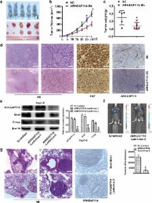
          ARHGAP11A exhibited high expression in HCC, and was significantly correlated with clinicopathological stage and prognosis in HCC patients. Moreover, ARHGAP11A facilitated Hep3B and MHCC97-H cell proliferation, invasion, migration and EMT development in vitro. ARHGAP11A knockdown significantly inhibited the in vivo growth and metastasis of HCC cells. Furthermore, ARHGAP11A directly interacted with Rac1B independent of Rho GTPase- activating activity. Rac1B blockade effectively interrupted ARHGAP11A-elicited HCC malignant phenotype. Meanwhile, upregulation of Rac1B reversed ARHGAP11A knockdown mediated mesenchymal-to-epithelial transition (MET) development in HCC cells.

          Conclusion

          ARHGAP11A facilitates malignant progression in HCC patients via ARHGAP11A-Rac1B interaction. The ARHGAP11A/Rac1B signaling could be a potential therapeutic target in the clinical treatment of HCC.

          Electronic supplementary material

          The online version of this article (10.1186/s12964-018-0312-4) contains supplementary material, which is available to authorized users.

          Related collections

          Most cited references33

          • Record: found
          • Abstract: found
          • Article: not found

          New signals from the invasive front.

          Approximately 90% of all cancer deaths arise from the metastatic spread of primary tumours. Of all the processes involved in carcinogenesis, local invasion and the formation of metastases are clinically the most relevant, but they are the least well understood at the molecular level. Revealing their mechanisms is one of the main challenges for exploratory and applied cancer research. Recent experimental progress has identified a number of molecular pathways and cellular mechanisms that underlie the multistage process of metastasis formation: these include tumour invasion, tumour-cell dissemination through the bloodstream or the lymphatic system, colonization of distant organs and, finally, fatal outgrowth of metastases.
            Bookmark
            • Record: found
            • Abstract: found
            • Article: not found

            Rac1b, a tumor associated, constitutively active Rac1 splice variant, promotes cellular transformation.

            A novel splice variant of Rac1, designated Rac1b, is expressed in human breast and colon carcinoma cells. Rac1b contains an additional 19 amino-acid insert immediately behind the switch II domain, a region important for Rac1 interaction with regulators and effectors. Recent studies showed that Rac1b exhibited the biochemical properties of a constitutively activated GTPase, yet it showed impaired interaction with downstream effectors, suggesting that Rac1b may be defective in biological activity. Whether Rac1b is a biologically active protein was not addressed. Therefore, we evaluated the biochemical, signaling and growth-promoting properties of authentic Rac1b. Similar to previous observations, we found that Rac1b showed enhanced intrinsic guanine nucleotide exchange activity, impaired intrinsic GTPase activity, and failed to interact with RhoGDI. Surprisingly, we found that Rac1b, like the constitutively-activated and transforming Rac1(Q61L) mutant, promoted growth transformation of NIH3T3 cells. Rac1b-expressing cells also showed a loss of density-dependent and anchorage-dependent growth. Surprisingly, unlike activated Rac1(61L), Rac1b did not show enhanced activation of the nuclear factor kappaB (NF-kappaB) transcription factor or stimulate cyclin D1 expression, the signaling activities that best correlate with Rac1 transforming activity. However, Rac1b did promote activation of the AKT serine/threonine kinase. Therefore, we suggest that Rac1b selectively activates a subset of Rac1 downstream signaling pathways to facilitate cellular transformation.
              Bookmark
              • Record: found
              • Abstract: found
              • Article: not found

              Cloning of a novel human Rac1b splice variant with increased expression in colorectal tumors.

              Rac1 is a member of the Ras superfamily of small GTPases involved in signal transduction pathways that induce the formation of lamellipodia, stimulate cell proliferation and activate the JNK/SAPK protein kinase cascade. Here we describe that amplification by RT-PCR of the entire Rac1 coding sequence from a series of human adult and fetal tissues revealed beside the expected Rac1 cDNA, a variant product which contained additional 57 nucleotides between codons 75 and 76. This variant resulted in an in-frame insertion of 19 new amino acids immediately behind the switch II region, including two potential threonine phosphorylation sites for casein kinase II and protein kinase C. Primers designed within and downstream of the inserted nucleotide sequence allowed isolation of a genomic clone with intronic consensus sequences demonstrating that the insertion corresponds to a novel, yet undescribed exon 3b. This Rac1 splice variant, designated Rac1b, was predominantly identified in skin and epithelial tissues from the intestinal tract. Most notably, the expression of rac1b versus rac1 was found to be elevated in colorectal tumors at various stages of neoplastic progression, as compared to their respective adjacent tissues. We suggest that the 19 amino acid-insertion following the switch II region may create a novel effector binding site in rac1b, and thus participate in signaling pathways related to the normal or neoplastic growth of the intestinal mucosa.
                Bookmark

                Author and article information

                Contributors
                drdbmd@163.com
                zhangxuanxj@163.com
                shangrunze@126.com
                wangjianlin217@163.com
                yangxisheng205@163.com
                ahrhong@126.com
                liuqwhu@fmmu.edu.cn
                wangdesh@163.com
                Fierywang@163.com
                doukef@fmmu.edu.cn
                Journal
                Cell Commun Signal
                Cell Commun. Signal
                Cell Communication and Signaling : CCS
                BioMed Central (London )
                1478-811X
                13 December 2018
                13 December 2018
                2018
                : 16
                : 99
                Affiliations
                ISNI 0000 0004 1799 374X, GRID grid.417295.c, Department of Hepatobiliary Surgery, , Xijing Hospital, The Fourth Military Medical University, ; Xi’an, China
                Article
                312
                10.1186/s12964-018-0312-4
                6293628
                30545369
                97dd809e-8032-4ae3-86fb-46155e445a92
                © The Author(s). 2018

                Open AccessThis article is distributed under the terms of the Creative Commons Attribution 4.0 International License ( http://creativecommons.org/licenses/by/4.0/), which permits unrestricted use, distribution, and reproduction in any medium, provided you give appropriate credit to the original author(s) and the source, provide a link to the Creative Commons license, and indicate if changes were made. The Creative Commons Public Domain Dedication waiver ( http://creativecommons.org/publicdomain/zero/1.0/) applies to the data made available in this article, unless otherwise stated.

                History
                : 24 July 2018
                : 3 December 2018
                Funding
                Funded by: FundRef http://dx.doi.org/10.13039/501100001809, National Natural Science Foundation of China;
                Award ID: 81672341
                Award Recipient :
                Categories
                Research
                Custom metadata
                © The Author(s) 2018

                Cell biology
                arhgap11a,hepatocellular carcinoma,emt,metastasis,rac1b
                Cell biology
                arhgap11a, hepatocellular carcinoma, emt, metastasis, rac1b

                Comments

                Comment on this article