3
views
0
recommends
+1 Recommend
0 collections
    0
    shares
      • Record: found
      • Abstract: found
      • Article: found
      Is Open Access

      Injection with Toxoplasma gondii protein affects neuron health and survival

      research-article

      Read this article at

      Bookmark
          There is no author summary for this article yet. Authors can add summaries to their articles on ScienceOpen to make them more accessible to a non-specialist audience.

          Abstract

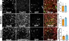
          Toxoplasma gondii is an intracellular parasite that causes a long-term latent infection of neurons. Using a custom MATLAB-based mapping program in combination with a mouse model that allows us to permanently mark neurons injected with parasite proteins, we found that Toxoplasma-injected neurons (TINs) are heterogeneously distributed in the brain, primarily localizing to the cortex followed by the striatum. In addition, we determined that cortical TINs are commonly (>50%) excitatory neurons (FoxP2 +) and that striatal TINs are often (>65%) medium spiny neurons (MSNs) (FoxP2 +). By performing single neuron patch clamping on striatal TINs and neighboring uninfected MSNs, we discovered that TINs have highly aberrant electrophysiology. As approximately 90% of TINs will die by 8 weeks post-infection, this abnormal physiology suggests that injection with Toxoplasma protein—either directly or indirectly—affects neuronal health and survival. Collectively, these data offer the first insights into which neurons interact with Toxoplasma and how these interactions alter neuron physiology in vivo.

          eLife digest

          Toxoplasma gondii is an intracellular parasite that infects the brain. Whereas most microbial infections of the brain result in severe illness or death, Toxoplasma gondii infections are usually asymptomatic. This is because the parasite has evolved the ability to exist within the brain by dampening the immune response. The parasite can therefore asymptomatically co-exist with its host for years – or even an entire lifetime. The strategy has proved so successful that up to one third of the world’s population is now thought to be infected with Toxoplasma gondii.

          While this persistence tends not to be a problem for most healthy individuals, dormant Toxoplasma gondii parasites can reactivate in individuals whose immune systems fail. This can result in life-threatening neurological disease. In pregnant women, Toxoplasma gondii parasites can also cross the placenta, which can trigger miscarriage or cause harmful disease in the newborn.

          To develop treatments for these cases of symptomatic disease, we need to understand how the parasite hides from the immune system in asymptomatic individuals. Mendez et al. have therefore leveraged a mouse model in which neurons injected with Toxoplasma gondii proteins ( Toxoplasma-injected neurons, or ‘TINs’) produce a green fluorescent protein. This enables the infected cells to be viewed under a microscope.

          Examining the mouse brains revealed that most TINs were located in two specific regions: the cortex and the striatum. The cortex is the brain’s outer layer of tissue. The striatum is a structure deep within the brain that helps regulate movement and responses to rewards. Both the cortex and the striatum contain different types of neurons. The results revealed that the proteins from the parasite were spread roughly equally among the various cell types, rather than targeting a specific subtype of neuron.

          Neurons close to TINs had slightly abnormal electrical activity, whereas the TINs themselves had highly abnormal activity. By eight weeks post-infection, however, the number of TINS had fallen by around 90%. This suggests that many neurons containing Toxoplasma protein are sick and dying, and that their altered electrical activity reflects this unhealthy state.

          Understanding how Toxoplasma parasites persist in the brain has the potential to reveal new targets for treating symptomatic infections. It could even provide new possibilities for targeting the inflammation that drives many other neurological diseases. Harnessing this potential will require finding out why Toxoplasma gondii infects specific brain regions and why most neurons that directly interact with the parasite die.

          Related collections

          Most cited references69

          • Record: found
          • Abstract: found
          • Article: not found

          A robust and high-throughput Cre reporting and characterization system for the whole mouse brain

          The Cre/lox system is widely used in mice to achieve cell-type-specific gene expression. However, a strong and universal responding system to express genes under Cre control is still lacking. We have generated a set of Cre reporter mice with strong, ubiquitous expression of fluorescent proteins of different spectra. The robust native fluorescence of these reporters enables direct visualization of fine dendritic structures and axonal projections of the labeled neurons, which is useful in mapping neuronal circuitry, imaging and tracking specific cell populations in vivo. Using these reporters and a high-throughput in situ hybridization platform, we are systematically profiling Cre-directed gene expression throughout the mouse brain in a number of Cre-driver lines, including novel Cre lines targeting different cell types in the cortex. Our expression data are displayed in a public online database to help researchers assess the utility of various Cre-driver lines for cell-type-specific genetic manipulation.
            Bookmark
            • Record: found
            • Abstract: found
            • Article: not found

            GABAergic Interneurons in the Neocortex: From Cellular Properties to Circuits.

            Cortical networks are composed of glutamatergic excitatory projection neurons and local GABAergic inhibitory interneurons that gate signal flow and sculpt network dynamics. Although they represent a minority of the total neocortical neuronal population, GABAergic interneurons are highly heterogeneous, forming functional classes based on their morphological, electrophysiological, and molecular features, as well as connectivity and in vivo patterns of activity. Here we review our current understanding of neocortical interneuron diversity and the properties that distinguish cell types. We then discuss how the involvement of multiple cell types, each with a specific set of cellular properties, plays a crucial role in diversifying and increasing the computational power of a relatively small number of simple circuit motifs forming cortical networks. We illustrate how recent advances in the field have shed light onto the mechanisms by which GABAergic inhibition contributes to network operations.
              Bookmark
              • Record: found
              • Abstract: found
              • Article: not found

              Neuronal subtype-specific genes that control corticospinal motor neuron development in vivo.

              Within the vertebrate nervous system, the presence of many different lineages of neurons and glia complicates the molecular characterization of single neuronal populations. In order to elucidate molecular mechanisms underlying the specification and development of corticospinal motor neurons (CSMN), we purified CSMN at distinct stages of development in vivo and compared their gene expression to two other pure populations of cortical projection neurons: callosal projection neurons and corticotectal projection neurons. We found genes that are potentially instructive for CSMN development, as well as genes that are excluded from CSMN and are restricted to other populations of neurons, even within the same cortical layer. Loss-of-function experiments in null mutant mice for Ctip2 (also known as Bcl11b), one of the newly characterized genes, demonstrate that it plays a critical role in the development of CSMN axonal projections to the spinal cord in vivo, confirming that we identified central genetic determinants of the CSMN population.
                Bookmark

                Author and article information

                Contributors
                Role: Reviewing Editor
                Role: Senior Editor
                Journal
                eLife
                Elife
                eLife
                eLife
                eLife Sciences Publications, Ltd
                2050-084X
                09 June 2021
                2021
                : 10
                : e67681
                Affiliations
                [1 ]Graduate Interdisciplinary Program in Neuroscience, University of Arizona TucsonUnited States
                [2 ]BIO5 Institute, University of Arizona TucsonUnited States
                [3 ]College of Nursing, University of Arizona TucsonUnited States
                [4 ]Department of Immunobiology, University of Arizona TucsonUnited States
                [5 ]Department of Neurology, University of Arizona TucsonUnited States
                The University of Melbourne Australia
                University of the Witwatersrand South Africa
                The University of Melbourne Australia
                Author information
                https://orcid.org/0000-0001-8705-3233
                Article
                67681
                10.7554/eLife.67681
                8270641
                34106047
                98122ea3-aada-493d-bf3e-96aeeb8af604
                © 2021, Mendez et al

                This article is distributed under the terms of the Creative Commons Attribution License, which permits unrestricted use and redistribution provided that the original author and source are credited.

                History
                : 18 February 2021
                : 09 June 2021
                Funding
                Funded by: FundRef http://dx.doi.org/10.13039/100000065, National Institute of Neurological Disorders and Stroke;
                Award ID: F99 NS108514
                Award Recipient :
                Funded by: FundRef http://dx.doi.org/10.13039/100000065, National Institute of Neurological Disorders and Stroke;
                Award ID: R25 NS076437
                Award Recipient :
                Funded by: FundRef http://dx.doi.org/10.13039/100008335, Arizona Biomedical Research Commission;
                Award ID: ADHS14-082991
                Award Recipient :
                Funded by: FundRef http://dx.doi.org/10.13039/100000065, National Institute of Neurological Disorders and Stroke;
                Award ID: R01 NS095994
                Award Recipient :
                The funders had no role in study design, data collection and interpretation, or the decision to submit the work for publication.
                Categories
                Research Article
                Microbiology and Infectious Disease
                Neuroscience
                Custom metadata
                Multi-level profiling of Toxoplasma gondii-injected neurons identifies brain regions and neuron subtype predilections as well as the effect of Toxoplasma interactions on neuron physiology and health.

                Life sciences
                toxoplasma gondii,electrophysiology,localization,mouse
                Life sciences
                toxoplasma gondii, electrophysiology, localization, mouse

                Comments

                Comment on this article