8
views
0
recommends
+1 Recommend
0 collections
    0
    shares
      • Record: found
      • Abstract: found
      • Article: not found

      Ankyrin-B Is Required for Intracellular Sorting of Structurally Diverse Ca 2+ Homeostasis Proteins

      research-article

      Read this article at

      ScienceOpenPMC
      Bookmark
          There is no author summary for this article yet. Authors can add summaries to their articles on ScienceOpen to make them more accessible to a non-specialist audience.

          Abstract

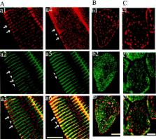
          This report describes a congenital myopathy and major loss of thymic lymphocytes in ankyrin-B (−/−) mice as well as dramatic alterations in intracellular localization of key components of the Ca 2+ homeostasis machinery in ankyrin-B (−/−) striated muscle and thymus. The sacoplasmic reticulum (SR) and SR/T-tubule junctions are apparently preserved in a normal distribution in ankyrin-B (−/−) skeletal muscle based on electron microscopy and the presence of a normal pattern of triadin and dihydropyridine receptor. Therefore, the abnormal localization of SR/ER Ca ATPase (SERCA) and ryanodine receptors represents a defect in intracellular sorting of these proteins in skeletal muscle. Extrapolation of these observations suggests defective targeting as the basis for abnormal localization of ryanodine receptors, IP3 receptors and SERCA in heart, and of IP3 receptors in the thymus of ankyrin-B (−/−) mice. Mis-sorting of SERCA 2 and ryanodine receptor 2 in ankyrin-B (−/−) cardiomyocytes is rescued by expression of 220-kD ankyrin-B, demonstrating that lack of the 220-kD ankyrin-B polypeptide is the primary defect in these cells. Ankyrin-B is associated with intracellular vesicles, but is not colocalized with the bulk of SERCA 1 or ryanodine receptor type 1 in skeletal muscle. These data provide the first evidence of a physiological requirement for ankyrin-B in intracellular targeting of the calcium homeostasis machinery of striated muscle and immune system, and moreover, support a catalytic role that does not involve permanent stoichiometric complexes between ankyrin-B and targeted proteins. Ankyrin-B is a member of a family of adapter proteins implicated in restriction of diverse proteins to specialized plasma membrane domains. Similar mechanisms involving ankyrins may be essential for segregation of functionally defined proteins within specialized regions of the plasma membrane and within the Ca 2+ homeostasis compartment of the ER.

          Related collections

          Most cited references40

          • Record: found
          • Abstract: found
          • Article: not found

          AnkyrinG Is Required for Clustering of Voltage-gated Na Channels at Axon Initial Segments and for Normal Action Potential Firing

          Voltage-gated sodium channels (NaCh) are colocalized with isoforms of the membrane-skeletal protein ankyrinG at axon initial segments, nodes of Ranvier, and postsynaptic folds of the mammalian neuromuscular junction. The role of ankyrinG in directing NaCh localization to axon initial segments was evaluated by region-specific knockout of ankyrinG in the mouse cerebellum. Mutant mice exhibited a progressive ataxia beginning around postnatal day P16 and subsequent loss of Purkinje neurons. In mutant mouse cerebella, NaCh were absent from axon initial segments of granule cell neurons, and Purkinje cells showed deficiencies in their ability to initiate action potentials and support rapid, repetitive firing. Neurofascin, a member of the L1CAM family of ankyrin-binding cell adhesion molecules, also exhibited impaired localization to initial segments of Purkinje cell neurons. These results demonstrate that ankyrinG is essential for clustering NaCh and neurofascin at axon initial segments and is required for physiological levels of sodium channel activity.
            Bookmark
            • Record: found
            • Abstract: found
            • Article: not found

            Ryanodine receptors of striated muscles: a complex channel capable of multiple interactions.

            The ryanodine receptor (RyR) is a high-conductance Ca2+ channel of the sarcoplasmic reticulum in muscle and of the endoplasmic reticulum in other cells. In striated muscle fibers, RyRs are responsible for the rapid release of Ca2+ that activates contraction. Ryanodine receptors are complex molecules, with unusually large cytoplasmic domains containing numerous binding sites for agents that control the state of activity of the channel-forming domain of the molecule. Structural considerations indicate that long-range interactions between cytoplasmic and intramembrane domains control channel function. Ryanodine receptors are located in specialized regions of the SR, where they are structurally and functionally associated with other intrinsic proteins and, indirectly, also with the luminal Ca2(+)-binding protein calsequestrin. Activation of RyRs during the early part of the excitation-contraction coupling cascade is initiated by the activity of surface-membrane Ca2+ channels, the dihydropyridine receptors (DHPRs). Skeletal and cardiac muscles contain different RyR and DHPR isoforms and both contribute to the diversity in cardiac and skeletal excitation-contraction coupling mechanisms. The architecture of the sarcoplasmic reticulum-surface junctions determines the types of RyR-DHPR interactions in the two muscle types.
              Bookmark
              • Record: found
              • Abstract: found
              • Article: not found

              Regulation of calcium signalling in T lymphocytes by the second messenger cyclic ADP-ribose.

              Cyclic ADP-ribose (cADPR) is a natural compound that mobilizes calcium ions in several eukaryotic cells. Although it can lead to the release of calcium ions in T lymphocytes, it has not been firmly established as a second messenger in these cells. Here, using high-performance liquid chromatography analysis, we show that stimulation of the T-cell receptor/CD3 (TCR/CD3) complex results in activation of a soluble ADP-ribosyl cyclase and a sustained increase in intracellular levels of cADPR. There is a causal relation between increased cADPR concentrations, sustained calcium signalling and activation of T cells, as shown by inhibition of TCR/CD3-stimulated calcium signalling, cell proliferation and expression of the early- and late-activation markers CD25 and HLA-DR by using cADPR antagonists. The molecular target for cADPR, the type-3 ryanodine receptor/calcium channel, is expressed in T cells. Increased cADPR significantly and specifically stimulates the apparent association of [3H]ryanodine with the type-3 ryanodine receptor, indicating a direct modulatory effect of cADPR on channel opening. Thus we show the presence, causal relation and biological significance of the major constituents of the cADPR/calcium-signalling pathway in human T cells.
                Bookmark

                Author and article information

                Contributors
                Journal
                J Cell Biol
                The Journal of Cell Biology
                The Rockefeller University Press
                0021-9525
                1540-8140
                29 November 1999
                : 147
                : 5
                : 995-1008
                Affiliations
                [a ]Howard Hughes Medical Institute and Departments of Cell Biology and Biochemistry, Duke University Medical Center, Durham, North Carolina 27710
                Article
                9906142
                2169334
                10579720
                a1399cd8-37bb-452f-8eca-558bd9662cdc
                © 1999 The Rockefeller University Press
                History
                : 30 June 1999
                : 21 September 1999
                : 20 October 1999
                Categories
                Original Article

                Cell biology
                calcium homeostasis,ip3 receptor,ankyrin,sarcoplasmic reticulum,gene knockout
                Cell biology
                calcium homeostasis, ip3 receptor, ankyrin, sarcoplasmic reticulum, gene knockout

                Comments

                Comment on this article