4
views
0
recommends
+1 Recommend
0 collections
    0
    shares
      • Record: found
      • Abstract: found
      • Article: found
      Is Open Access

      Physical exercise promotes astrocyte coverage of microvessels in a model of chronic cerebral hypoperfusion

      research-article

      Read this article at

      Bookmark
          There is no author summary for this article yet. Authors can add summaries to their articles on ScienceOpen to make them more accessible to a non-specialist audience.

          Abstract

          Background

          Brain circulation disorders such as chronic cerebral hypoperfusion have been associated with a decline in cognitive function during the development of dementia. Astrocytes together with microglia participate in the immune response in the CNS and make them potential sentinels in the brain parenchyma. In addition, astrocytes coverage integrity has been related to brain homeostasis. Currently, physical exercise has been proposed as an effective intervention to promote brain function improvement. However, the neuroprotective effects of early physical exercise on the astrocyte communication with the microcirculation and the microglial activation in a chronic cerebral hypoperfusion model are still unclear. The aim of this study was to investigate the impact of early intervention with physical exercise on cognition, brain microcirculatory, and inflammatory parameters in an experimental model of chronic cerebral hypoperfusion induced by permanent bilateral occlusion of the common carotid arteries (2VO).

          Methods

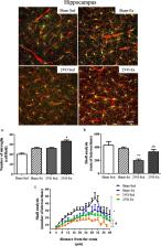
          Wistar rats aged 12 weeks were randomly divided into four groups: Sham-sedentary group (Sham-Sed), Sham-exercised group (Sham-Ex), 2VO-sedentary group (2VO-Sed), and 2VO-exercised group (2VO-Ex). The early intervention with physical exercise started 3 days after 2VO or Sham surgery during 12 weeks. Then, the brain functional capillary density and endothelial-leukocyte interactions were evaluated by intravital microscopy; cognitive function was evaluated by open-field test; hippocampus postsynaptic density protein 95 and synaptophysin were evaluated by western blotting; astrocytic coverage of the capillaries, microglial activation, and structural capillary density were evaluated by immunohistochemistry.

          Results

          Early moderate physical exercise was able to normalize functional capillary density and reduce leukocyte rolling in the brain of animals with chronic cerebral hypoperfusion. These effects were accompanied by restore synaptic protein and the improvement of cognitive function. In addition, early moderate exercise improves astrocytes coverage in blood vessels of the cerebral cortex and hippocampus, decreases microglial activation in the hippocampus, and improves structural capillaries in the hippocampus.

          Conclusions

          Microcirculatory and inflammatory changes in the brain appear to be involved in triggering a cognitive decline in animals with chronic cerebral ischemia. Therefore, early intervention with physical exercise may represent a preventive approach to neurodegeneration caused by chronic cerebral hypoperfusion.

          Related collections

          Most cited references53

          • Record: found
          • Abstract: found
          • Article: not found

          Cardiovascular fitness, cortical plasticity, and aging.

          Cardiovascular fitness is thought to offset declines in cognitive performance, but little is known about the cortical mechanisms that underlie these changes in humans. Research using animal models shows that aerobic training increases cortical capillary supplies, the number of synaptic connections, and the development of new neurons. The end result is a brain that is more efficient, plastic, and adaptive, which translates into better performance in aging animals. Here, in two separate experiments, we demonstrate for the first time to our knowledge, in humans that increases in cardiovascular fitness results in increased functioning of key aspects of the attentional network of the brain during a cognitively challenging task. Specifically, highly fit (Study 1) or aerobically trained (Study 2) persons show greater task-related activity in regions of the prefrontal and parietal cortices that are involved in spatial selection and inhibitory functioning, when compared with low-fit (Study 1) or nonaerobic control (Study 2) participants. Additionally, in both studies there exist groupwise differences in activation of the anterior cingulate cortex, which is thought to monitor for conflict in the attentional system, and signal the need for adaptation in the attentional network. These data suggest that increased cardiovascular fitness can affect improvements in the plasticity of the aging human brain, and may serve to reduce both biological and cognitive senescence in humans.
            Bookmark
            • Record: found
            • Abstract: found
            • Article: not found

            Interplay of hippocampus and prefrontal cortex in memory.

            Recent studies on the hippocampus and the prefrontal cortex have considerably advanced our understanding of the distinct roles of these brain areas in the encoding and retrieval of memories, and of how they interact in the prolonged process by which new memories are consolidated into our permanent storehouse of knowledge. These studies have led to a new model of how the hippocampus forms and replays memories and how the prefrontal cortex engages representations of the meaningful contexts in which related memories occur, as well as how these areas interact during memory retrieval. Furthermore, they have provided new insights into how interactions between the hippocampus and prefrontal cortex support the assimilation of new memories into pre-existing networks of knowledge, called schemas, and how schemas are modified in this process as the foundation of memory consolidation. Copyright © 2013 Elsevier Ltd. All rights reserved.
              Bookmark
              • Record: found
              • Abstract: found
              • Article: found
              Is Open Access

              A Computational Tool for Quantitative Analysis of Vascular Networks

              Angiogenesis is the generation of mature vascular networks from pre-existing vessels. Angiogenesis is crucial during the organism' development, for wound healing and for the female reproductive cycle. Several murine experimental systems are well suited for studying developmental and pathological angiogenesis. They include the embryonic hindbrain, the post-natal retina and allantois explants. In these systems vascular networks are visualised by appropriate staining procedures followed by microscopical analysis. Nevertheless, quantitative assessment of angiogenesis is hampered by the lack of readily available, standardized metrics and software analysis tools. Non-automated protocols are being used widely and they are, in general, time - and labour intensive, prone to human error and do not permit computation of complex spatial metrics. We have developed a light-weight, user friendly software, AngioTool, which allows for quick, hands-off and reproducible quantification of vascular networks in microscopic images. AngioTool computes several morphological and spatial parameters including the area covered by a vascular network, the number of vessels, vessel length, vascular density and lacunarity. In addition, AngioTool calculates the so-called “branching index” (branch points / unit area), providing a measurement of the sprouting activity of a specimen of interest. We have validated AngioTool using images of embryonic murine hindbrains, post-natal retinas and allantois explants. AngioTool is open source and can be downloaded free of charge.
                Bookmark

                Author and article information

                Contributors
                estato@ioc.fiocruz.br , vanessaestato@gmail.com
                Journal
                J Neuroinflammation
                J Neuroinflammation
                Journal of Neuroinflammation
                BioMed Central (London )
                1742-2094
                16 April 2020
                16 April 2020
                2020
                : 17
                : 117
                Affiliations
                [1 ]GRID grid.418068.3, ISNI 0000 0001 0723 0931, Laboratory of Immunopharmacology, , Oswaldo Cruz Foundation, ; Av. Brasil, 4365, Manguinhos, Rio de Janeiro, 21040-900 Brazil
                [2 ]GRID grid.418068.3, ISNI 0000 0001 0723 0931, Laboratory of Cardiovascular Investigation, , Oswaldo Cruz Foundation, ; Rio de Janeiro, Brazil
                [3 ]GRID grid.8536.8, ISNI 0000 0001 2294 473X, Laboratory of Glial Cell Biology, Biomedical Sciences Institute, , Federal University of Rio de Janeiro, ; Rio de Janeiro, Brazil
                [4 ]GRID grid.419171.b, ISNI 0000 0004 0481 7106, National Institute of Cardiology, ; Rio de Janeiro, Brazil
                Article
                1771
                10.1186/s12974-020-01771-y
                7161182
                32299450
                a1a5b113-b5b8-4fde-b530-b28394b34695
                © The Author(s) 2020

                Open AccessThis article is licensed under a Creative Commons Attribution 4.0 International License, which permits use, sharing, adaptation, distribution and reproduction in any medium or format, as long as you give appropriate credit to the original author(s) and the source, provide a link to the Creative Commons licence, and indicate if changes were made. The images or other third party material in this article are included in the article's Creative Commons licence, unless indicated otherwise in a credit line to the material. If material is not included in the article's Creative Commons licence and your intended use is not permitted by statutory regulation or exceeds the permitted use, you will need to obtain permission directly from the copyright holder. To view a copy of this licence, visit http://creativecommons.org/licenses/by/4.0/. The Creative Commons Public Domain Dedication waiver ( http://creativecommons.org/publicdomain/zero/1.0/) applies to the data made available in this article, unless otherwise stated in a credit line to the data.

                History
                : 6 January 2020
                : 12 March 2020
                Funding
                Funded by: FundRef http://dx.doi.org/10.13039/501100003593, Conselho Nacional de Desenvolvimento Científico e Tecnológico;
                Funded by: Fundação de Amparo à Pesquisa do Estado do Rio de Janeiro
                Funded by: FundRef http://dx.doi.org/10.13039/501100006507, Fundação Oswaldo Cruz;
                Categories
                Research
                Custom metadata
                © The Author(s) 2020

                Neurosciences
                exercise,cerebral hypoperfusion,neuroinflammation,glial cells,microcirculation
                Neurosciences
                exercise, cerebral hypoperfusion, neuroinflammation, glial cells, microcirculation

                Comments

                Comment on this article