57
views
0
recommends
+1 Recommend
0 collections
    0
    shares
      • Record: found
      • Abstract: found
      • Article: not found

      Abnormal accumulation of autophagic vesicles correlates with axonal and synaptic pathology in young Alzheimer’s mice hippocampus

      research-article

      Read this article at

      ScienceOpenPublisherPMC
      Bookmark
          There is no author summary for this article yet. Authors can add summaries to their articles on ScienceOpen to make them more accessible to a non-specialist audience.

          Abstract

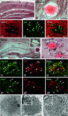
          Dystrophic neurites associated with amyloid plaques precede neuronal death and manifest early in Alzheimer’s disease (AD). In this work we have characterized the plaque-associated neuritic pathology in the hippocampus of young (4- to 6-month-old) PS1 M146L/APP 751SL mice model, as the initial degenerative process underlying functional disturbance prior to neuronal loss. Neuritic plaques accounted for almost all fibrillar deposits and an axonal origin of the dystrophies was demonstrated. The early induction of autophagy pathology was evidenced by increased protein levels of the autophagosome marker LC3 that was localized in the axonal dystrophies, and by electron microscopic identification of numerous autophagic vesicles filling and causing the axonal swellings. Early neuritic cytoskeletal defects determined by the presence of phosphorylated tau (AT8-positive) and actin–cofilin rods along with decreased levels of kinesin-1 and dynein motor proteins could be responsible for this extensive vesicle accumulation within dystrophic neurites. Although microsomal Aβ oligomers were identified, the presence of A11-immunopositive Aβ plaques also suggested a direct role of plaque-associated Aβ oligomers in defective axonal transport and disease progression. Most importantly, presynaptic terminals morphologically disrupted by abnormal autophagic vesicle buildup were identified ultrastructurally and further supported by synaptosome isolation. Finally, these early abnormalities in axonal and presynaptic structures might represent the morphological substrate of hippocampal dysfunction preceding synaptic and neuronal loss and could significantly contribute to AD pathology in the preclinical stages.

          Related collections

          Most cited references38

          • Record: found
          • Abstract: found
          • Article: not found

          Axonopathy and transport deficits early in the pathogenesis of Alzheimer's disease.

          We identified axonal defects in mouse models of Alzheimer's disease that preceded known disease-related pathology by more than a year; we observed similar axonal defects in the early stages of Alzheimer's disease in humans. Axonal defects consisted of swellings that accumulated abnormal amounts of microtubule-associated and molecular motor proteins, organelles, and vesicles. Impairing axonal transport by reducing the dosage of a kinesin molecular motor protein enhanced the frequency of axonal defects and increased amyloid-beta peptide levels and amyloid deposition. Reductions in microtubule-dependent transport may stimulate proteolytic processing of beta-amyloid precursor protein, resulting in the development of senile plaques and Alzheimer's disease.
            Bookmark
            • Record: found
            • Abstract: found
            • Article: not found

            Synaptic alterations in CA1 in mild Alzheimer disease and mild cognitive impairment.

            To evaluate the total number of synapses in the stratum radiatum (str rad) of the human hippocampal CA1 subfield in individuals with mild Alzheimer disease (mAD), mild cognitive impairment (MCI), or no cognitive impairment (NCI) and determine if synapse loss is an early event in the progression of the disease. Short postmortem autopsy tissue was obtained, and an unbiased stereologic sampling scheme coupled with transmission electron microscopy was used to directly visualize synaptic contacts. Individuals with mAD had fewer synapses (55%) than the other two diagnostic groups. Individuals with MCI had a mean synaptic value that was 18% lower than the NCI group mean. The total number of synapses showed a correlation with several cognitive tests including those involving both immediate and delayed recall. Total synaptic numbers showed no relationship to the subject's Braak stage or to APOE genotype. The volume of the str rad was reduced in mAD vs the other two diagnostic groups that were not different from each other. These results strongly support the concept that synapse loss is a structural correlate involved very early in cognitive decline in mild Alzheimer disease (mAD) and supports mild cognitive impairment as a transitional stage between mAD and no cognitive impairment.
              Bookmark
              • Record: found
              • Abstract: found
              • Article: not found

              Endocytosis is required for synaptic activity-dependent release of amyloid-beta in vivo.

              Aggregation of amyloid-beta (Abeta) peptide into soluble and insoluble forms within the brain extracellular space is central to the pathogenesis of Alzheimer's disease. Full-length amyloid precursor protein (APP) is endocytosed from the cell surface into endosomes where it is cleaved to produce Abeta. Abeta is subsequently released into the brain interstitial fluid (ISF). We hypothesized that synaptic transmission results in more APP endocytosis, thereby increasing Abeta generation and release into the ISF. We found that inhibition of clathrin-mediated endocytosis immediately lowers ISF Abeta levels in vivo. Two distinct methods that increased synaptic transmission resulted in an elevation of ISF Abeta levels. Inhibition of endocytosis, however, prevented the activity-dependent increase in Abeta. We estimate that approximately 70% of ISF Abeta arises from endocytosis-associated mechanisms, with the vast majority of this pool also dependent on synaptic activity. These findings have implications for AD pathogenesis and may provide insights into therapeutic intervention.
                Bookmark

                Author and article information

                Contributors
                +34-952133344 , +34-952131937 , agutierrez@uma.es
                Journal
                Acta Neuropathol
                Acta Neuropathologica
                Springer-Verlag (Berlin/Heidelberg )
                0001-6322
                1432-0533
                22 October 2011
                22 October 2011
                January 2012
                : 123
                : 1
                : 53-70
                Affiliations
                [1 ]Dpto. Biología Celular, Genética y Fisiología, Facultad de Ciencias, Universidad de Málaga, Campus de Teatinos, 29071 Málaga, Spain
                [2 ]Dpto. Bioquímica y Biología Molecular, Facultad de Farmacia, Universidad de Sevilla, Seville, Spain
                [3 ]Laboratorio de Morfología Celular, Unidad Mixta Centro de Investigación Príncipe Felipe-UVEG, Valencia, Spain
                [4 ]Centro de Investigación Biomédica en Red sobre Enfermedades Neurodegenerativas (CIBERNED), Madrid, Spain
                [5 ]Instituto de Biomedicina de Sevilla (IBiS)-Hospital Universitario Virgen del Rocío/CSIC/Universidad de Sevilla, Seville, Spain
                Article
                896
                10.1007/s00401-011-0896-x
                3249205
                22020633
                a5d44508-debb-4a92-a980-9fcbe7390de4
                © The Author(s) 2011
                History
                : 22 July 2011
                : 27 September 2011
                : 13 October 2011
                Categories
                Original Paper
                Custom metadata
                © Springer-Verlag 2012

                Neurology
                presynaptic terminals,electron microscopy,ps1/app transgenic mice,lc3,dystrophic neurites,amyloid plaques

                Comments

                Comment on this article