4
views
0
recommends
+1 Recommend
0 collections
    0
    shares
      • Record: found
      • Abstract: found
      • Article: found
      Is Open Access

      Neuroanatomical and Microglial Alterations in the Striatum of Levodopa-Treated, Dyskinetic Hemi-Parkinsonian Rats

      research-article

      Read this article at

      Bookmark
          There is no author summary for this article yet. Authors can add summaries to their articles on ScienceOpen to make them more accessible to a non-specialist audience.

          Abstract

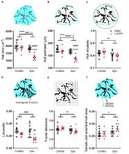
          Dyskinesia associated with chronic levodopa treatment in Parkinson’s disease is associated with maladaptive striatal plasticity. The objective of this study was to examine whether macroscale structural changes, as captured by magnetic resonance imaging (MRI) accompany this plasticity and to identify plausible cellular contributors in a rodent model of levodopa-induced dyskinesia. Adult male Sprague-Dawley rats were rendered hemi-parkinsonian by stereotaxic injection of 6-hydroxydopamine into the left medial forebrain bundle prior to chronic treatment with saline (control) or levodopa to induce abnormal involuntary movements (AIMs), reflective of dyskinesia. Perfusion-fixed brains underwent ex vivo structural MRI before sectioning and staining for cellular markers. Chronic treatment with levodopa induced significant AIMs ( p < 0.0001 versus saline). The absolute volume of the ipsilateral, lesioned striatum was increased in levodopa-treated rats resulting in a significant difference in percentage volume change when compared to saline-treated rats ( p < 0.01). Moreover, a significant positive correlation was found between this volume change and AIMs scores for individual levodopa-treated rats ( r = 0.96; p < 0.01). The density of Iba1+ cells was increased within the lesioned versus intact striatum ( p < 0.01) with no difference between treatment groups. Conversely, Iba1+ microglia soma size was significantly increased ( p < 0.01) in the lesioned striatum of levodopa-treated but not saline-treated rats. Soma size was not, however, significantly correlated with either AIMs or MRI volume change. Although GFAP+ astrocytes were elevated in the lesioned versus intact striatum ( p < 0.001), there was no difference between treatment groups. No statistically significant effects of either lesion or treatment on RECA1, a marker for blood vessels, were observed. Collectively, these data suggest chronic levodopa treatment in 6-hydroxydopamine lesioned rats is associated with increased striatal volume that correlates with the development of AIMs. The accompanying increase in number and size of microglia, however, cannot alone explain this volume expansion. Further multi-modal studies are warranted to establish the brain-wide effects of chronic levodopa treatment.

          Related collections

          Most cited references60

          • Record: found
          • Abstract: found
          • Article: found
          Is Open Access

          Quantitating the subtleties of microglial morphology with fractal analysis

          It is well established that microglial form and function are inextricably linked. In recent years, the traditional view that microglial form ranges between “ramified resting” and “activated amoeboid” has been emphasized through advancing imaging techniques that point to microglial form being highly dynamic even within the currently accepted morphological categories. Moreover, microglia adopt meaningful intermediate forms between categories, with considerable crossover in function and varying morphologies as they cycle, migrate, wave, phagocytose, and extend and retract fine and gross processes. From a quantitative perspective, it is problematic to measure such variability using traditional methods, but one way of quantitating such detail is through fractal analysis. The techniques of fractal analysis have been used for quantitating microglial morphology, to categorize gross differences but also to differentiate subtle differences (e.g., amongst ramified cells). Multifractal analysis in particular is one technique of fractal analysis that may be useful for identifying intermediate forms. Here we review current trends and methods of fractal analysis, focusing on box counting analysis, including lacunarity and multifractal analysis, as applied to microglial morphology.
            Bookmark
            • Record: found
            • Abstract: found
            • Article: not found

            Cell type-specific plasticity of striatal projection neurons in parkinsonism and L-DOPA-induced dyskinesia

            Summary The striatum is widely viewed as the fulcrum of pathophysiology in Parkinson's disease (PD) and L-DOPA-induced dyskinesia (LID). In these disease states, the balance in activity of striatal direct pathway spiny projection neurons (dSPNs) and indirect pathway spiny projection neurons (iSPNs) is disrupted, leading to aberrant action selection. However, it is unclear whether countervailing mechanisms are engaged in these states. Here we report that iSPN intrinsic excitability and excitatory corticostriatal synaptic connectivity were lower in PD models than normal; L-DOPA treatment restored these properties. Conversely, dSPN intrinsic excitability was elevated in tissue from PD models and suppressed in LID models. Although the synaptic connectivity of dSPNs did not change in PD models, it fell with L-DOPA treatment. In neither case, however, was the strength of corticostriatal connections globally scaled. Thus, SPNs manifested homeostatic adaptations in intrinsic excitability and in the number but not strength of excitatory corticostriatal synapses.
              Bookmark
              • Record: found
              • Abstract: found
              • Article: not found

              L-DOPA-induced dyskinesia in the rat is associated with striatal overexpression of prodynorphin- and glutamic acid decarboxylase mRNA.

              Rats sustaining unilateral near-complete 6-hydroxydopamine lesions of the mesostriatal dopamine pathway received daily injections of 3, 4 dihydroxyphenyl-l-alanine (L-DOPA, 8 mg/kg plus 15 mg/kg benserazide) for 3 weeks. During this period, about 50% of the rats gradually developed abnormal involuntary movements, lasting for 2-3 h following each L-DOPA dose. Rats were killed 3 days after the last L-DOPA injection, and sections through the striatum were processed for in situ hybridization histochemistry. Within the L-DOPA-treated group, levels of preproenkephalin (PPE) mRNA, glutamic acid decarboxylase (GAD67) mRNA, and prodynorphin (PDyn) mRNA in the dopamine-denervated caudate-putamen, as well as GAD67 mRNA expression in the globus pallidus ipsilateral to the 6-hydroxydopamine (6-OHDA) lesion, were higher in dyskinetic than non-dyskinetic animals, and positively correlated with the rats' dyskinesia scores. By contrast, striatal preprotachykinin mRNA expression and D2 receptor-radioligand binding were not significantly associated with dyskinesia. Among all these markers, PDyn mRNA levels showed the most pronounced treatment-dependence (three times higher in the L-DOPA-treated group than in saline-injected lesion-only controls), and the strongest correlation with the rats' dyskinesia scores (r2 = 0.82). However, a multiple regression equation including the three factors, GAD67 mRNA levels in the GP, GAD67 mRNA in the lateral CPu, and striatal PDyn mRNA, gave a better fit for dyskinesia scores than PDyn mRNA alone (r2 = 0.92). The results show that L-DOPA-induced dyskinesia is associated with overexpression of PDyn and GAD67 mRNA in the striatal projection neurons, and GAD67 mRNA levels in the globus pallidus. Due to its treatment-dependent expression, and strong correlation with the associated dyskinetic symptoms, striatal PDyn mRNA, in particular, may play a role in the mechanisms of behavioural sensitization brought about by the drug.
                Bookmark

                Author and article information

                Contributors
                Journal
                Front Neurosci
                Front. Neurosci.
                Frontiers in Neuroscience
                Frontiers Media S.A.
                1662-4548
                1662-453X
                15 September 2020
                2020
                : 14
                : 567222
                Affiliations
                [1] 1Wolfson Centre for Age Related Diseases, Wolfson Wing, Institute of Psychiatry, Psychology and Neuroscience, King’s College London , London, United Kingdom
                [2] 2Department of Neuroimaging, Institute of Psychiatry, Psychology and Neuroscience, King’s College London , London, United Kingdom
                [3] 3Department of Basic and Clinical Neuroscience, Maurice Wohl Clinical Neuroscience Institute, Institute of Psychiatry, Psychology and Neuroscience, King’s College London , London, United Kingdom
                [4] 4MRC Centre for Neurodevelopmental Disorders, King’s College London , London, United Kingdom
                Author notes

                Edited by: Tiago F. Outeiro, University Medical Center Goettingen, Germany

                Reviewed by: Erwan Bezard, UMR5293 Institut des Maladies Neurodégénératives (IMN), France; Rita Raisman-Vozari, Inserm, France; M. Angela Cenci, Lund University, Sweden

                *Correspondence: Susan Duty, susan.duty@ 123456kcl.ac.uk

                These authors share senior authorship

                This article was submitted to Neurodegeneration, a section of the journal Frontiers in Neuroscience

                Article
                10.3389/fnins.2020.567222
                7522511
                33041762
                a60b792d-f0f6-4d77-8173-a57df2725482
                Copyright © 2020 Fletcher, Finlay, Amor Lopez, Crum, Vernon and Duty.

                This is an open-access article distributed under the terms of the Creative Commons Attribution License (CC BY). The use, distribution or reproduction in other forums is permitted, provided the original author(s) and the copyright owner(s) are credited and that the original publication in this journal is cited, in accordance with accepted academic practice. No use, distribution or reproduction is permitted which does not comply with these terms.

                History
                : 29 May 2020
                : 17 August 2020
                Page count
                Figures: 5, Tables: 0, Equations: 0, References: 70, Pages: 15, Words: 0
                Funding
                Funded by: Medical Research Council 10.13039/501100000265
                Award ID: F67057A
                Award ID: G1002198
                Funded by: Guy's and St Thomas' Charity 10.13039/501100000380
                Award ID: R140805
                Categories
                Neuroscience
                Original Research

                Neurosciences
                abnormal involuntary movements,astrocytes,6-hydroxydopamine,levodopa-induced dyskinesia,magnetic resonance imaging,microglia,microvasculature

                Comments

                Comment on this article