63
views
0
recommends
+1 Recommend
0 collections
    0
    shares
      • Record: found
      • Abstract: found
      • Article: not found

      Atomic Resolution Insights into Curli Fiber Biogenesis

      research-article

      Read this article at

      ScienceOpenPublisherPMC
      Bookmark
          There is no author summary for this article yet. Authors can add summaries to their articles on ScienceOpen to make them more accessible to a non-specialist audience.

          Summary

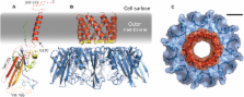
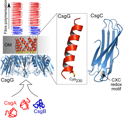
          Bacteria produce functional amyloid fibers called curli in a controlled, noncytotoxic manner. These extracellular fimbriae enable biofilm formation and promote pathogenicity. Understanding curli biogenesis is important for appreciating microbial lifestyles and will offer clues as to how disease-associated human amyloid formation might be ameliorated. Proteins encoded by the curli specific genes ( csgA-G) are required for curli production. We have determined the structure of CsgC and derived the first structural model of the outer-membrane subunit translocator CsgG. Unexpectedly, CsgC is related to the N-terminal domain of DsbD, both in structure and oxido-reductase capability. Furthermore, we show that CsgG belongs to the nascent class of helical outer-membrane macromolecular exporters. A cysteine in a CsgG transmembrane helix is a potential target of CsgC, and mutation of this residue influences curli assembly. Our study provides the first high-resolution structural insights into curli biogenesis.

          Abstract

          Graphical Abstract

          Highlights

          ► CsgC is related to the redox-active N-terminal domain of DsbD ► Outer-membrane transporter CsgG inserts into the membrane via an α-helical oligomer ► The transmembrane domain of CsgG is vital for pore forming and curli assembly ► CsgC appears to affect CsgG pore behavior and biofilm formation

          Related collections

          Most cited references47

          • Record: found
          • Abstract: found
          • Article: not found

          The PSIPRED protein structure prediction server.

          The PSIPRED protein structure prediction server allows users to submit a protein sequence, perform a prediction of their choice and receive the results of the prediction both textually via e-mail and graphically via the web. The user may select one of three prediction methods to apply to their sequence: PSIPRED, a highly accurate secondary structure prediction method; MEMSAT 2, a new version of a widely used transmembrane topology prediction method; or GenTHREADER, a sequence profile based fold recognition method. Freely available to non-commercial users at http://globin.bio.warwick.ac.uk/psipred/
            Bookmark
            • Record: found
            • Abstract: found
            • Article: not found

            REFMAC5 dictionary: organization of prior chemical knowledge and guidelines for its use.

            One of the most important aspects of macromolecular structure refinement is the use of prior chemical knowledge. Bond lengths, bond angles and other chemical properties are used in restrained refinement as subsidiary conditions. This contribution describes the organization and some aspects of the use of the flexible and human/machine-readable dictionary of prior chemical knowledge used by the maximum-likelihood macromolecular-refinement program REFMAC5. The dictionary stores information about monomers which represent the constitutive building blocks of biological macromolecules (amino acids, nucleic acids and saccharides) and about numerous organic/inorganic compounds commonly found in macromolecular crystallography. It also describes the modifications the building blocks undergo as a result of chemical reactions and the links required for polymer formation. More than 2000 monomer entries, 100 modification entries and 200 link entries are currently available. Algorithms and tools for updating and adding new entries to the dictionary have also been developed and are presented here. In many cases, the REFMAC5 dictionary allows entirely automatic generation of restraints within REFMAC5 refinement runs.
              Bookmark
              • Record: found
              • Abstract: found
              • Article: not found

              Protein structure alignment by incremental combinatorial extension (CE) of the optimal path.

              A new algorithm is reported which builds an alignment between two protein structures. The algorithm involves a combinatorial extension (CE) of an alignment path defined by aligned fragment pairs (AFPs) rather than the more conventional techniques using dynamic programming and Monte Carlo optimization. AFPs, as the name suggests, are pairs of fragments, one from each protein, which confer structure similarity. AFPs are based on local geometry, rather than global features such as orientation of secondary structures and overall topology. Combinations of AFPs that represent possible continuous alignment paths are selectively extended or discarded thereby leading to a single optimal alignment. The algorithm is fast and accurate in finding an optimal structure alignment and hence suitable for database scanning and detailed analysis of large protein families. The method has been tested and compared with results from Dali and VAST using a representative sample of similar structures. Several new structural similarities not detected by these other methods are reported. Specific one-on-one alignments and searches against all structures as found in the Protein Data Bank (PDB) can be performed via the Web at http://cl.sdsc.edu/ce.html.
                Bookmark

                Author and article information

                Journal
                Structure
                Structure
                Structure(London, England:1993)
                Cell Press
                0969-2126
                1878-4186
                07 September 2011
                07 September 2011
                : 19
                : 9-16
                : 1307-1316
                Affiliations
                [1 ]Division of Molecular Biosciences, Faculty of Natural Sciences, Imperial College London, London, SW7 2AZ, UK
                [2 ]Department of Molecular, Cellular, and Developmental Biology, University of Michigan, Ann Arbor, MI 48109, USA
                Author notes
                []Corresponding author s.j.matthews@ 123456imperial.ac.uk
                [∗∗ ]Corresponding author chapmanm@ 123456umich.edu
                [3]

                Present address: MRC National Institute for Medical Research, The Ridgeway, Mill Hill, London, NW7 1AA, UK

                [4]

                Present address: EMBL-EBI Hinxton, Cambridge, CB10 1SD, UK

                Article
                STFODE2238
                10.1016/j.str.2011.05.015
                3173608
                21893289
                abc3fd29-78c2-4453-9a2d-53ebedcab04b
                © 2011 ELL & Excerpta Medica.

                This document may be redistributed and reused, subject to certain conditions.

                History
                : 20 January 2011
                : 26 May 2011
                : 28 May 2011
                Categories
                Article

                Molecular biology
                Molecular biology

                Comments

                Comment on this article