33
views
0
recommends
+1 Recommend
0 collections
    0
    shares
      • Record: found
      • Abstract: found
      • Article: found
      Is Open Access

      Mutant Huntingtin induces activation of the Bcl-2/adenovirus E1B 19-kDa interacting protein (BNip3)

      research-article

      Read this article at

      Bookmark
          There is no author summary for this article yet. Authors can add summaries to their articles on ScienceOpen to make them more accessible to a non-specialist audience.

          Abstract

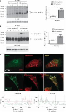
          Huntington's disease (HD) is a neurodegenerative disorder characterized by progressive neuronal death in the basal ganglia and cortex. Although increasing evidence supports a pivotal role of mitochondrial dysfunction in the death of patients' neurons, the molecular bases for mitochondrial impairment have not been elucidated. We provide the first evidence of an abnormal activation of the Bcl-2/adenovirus E1B 19-kDa interacting protein 3 (BNip3) in cells expressing mutant Huntingtin. In this study, we show an abnormal accumulation and dimerization of BNip3 in the mitochondria extracted from human HD muscle cells, HD model cell cultures and brain tissues from HD model mice. Importantly, we have shown that blocking BNip3 expression and dimerization restores normal mitochondrial potential in human HD muscle cells. Our data shed light on the molecular mechanisms underlying mitochondrial dysfunction in HD and point to BNip3 as a new potential target for neuroprotective therapy in HD.

          Related collections

          Most cited references31

          • Record: found
          • Abstract: found
          • Article: not found

          p53 mediates cellular dysfunction and behavioral abnormalities in Huntington's disease.

          We present evidence for a specific role of p53 in the mitochondria-associated cellular dysfunction and behavioral abnormalities of Huntington's disease (HD). Mutant huntingtin (mHtt) with expanded polyglutamine (polyQ) binds to p53 and upregulates levels of nuclear p53 as well as p53 transcriptional activity in neuronal cultures. The augmentation is specific, as it occurs with mHtt but not mutant ataxin-1 with expanded polyQ. p53 levels are also increased in the brains of mHtt transgenic (mHtt-Tg) mice and HD patients. Perturbation of p53 by pifithrin-alpha, RNA interference, or genetic deletion prevents mitochondrial membrane depolarization and cytotoxicity in HD cells, as well as the decreased respiratory complex IV activity of mHtt-Tg mice. Genetic deletion of p53 suppresses neurodegeneration in mHtt-Tg flies and neurobehavioral abnormalities of mHtt-Tg mice. Our findings suggest that p53 links nuclear and mitochondrial pathologies characteristic of HD.
            Bookmark
            • Record: found
            • Abstract: found
            • Article: not found

            Mitochondrial defect in Huntington's disease caudate nucleus.

            Although the Huntington's disease (HD) gene defect has been identified, the structure and function of the abnormal gene product and the pathogenetic mechanisms involved in producing death of selective neuronal populations are not understood. Indirect evidence from several sources indicates that a defect of energy metabolism and consequent excitotoxicity are involved in HD. Toxin models of HD may be induced by 3-nitropropionic acid or malonate, both inhibitors of succinate dehydrogenase, complex II of the mitochondrial respiratory chain. We analyzed mitochondrial respiratory chain function in the caudate nucleus (n = 10) and platelets (n = 11) from patients with HD. In the caudate nucleus, severe defects of complexes II and III (53-59%, p < 0.0005) and a 32-38% (p < 0.01) deficiency of complex IV activity were demonstrated. No deficiencies were found in platelet mitochondrial function. The mitochondrial defect identified in HD caudate parallels that induced by HD neurotoxin models and further supports the role of abnormal energy metabolism in HD. The relationship of the mitochondrial defect to the role of huntingtin is not known.
              Bookmark
              • Record: found
              • Abstract: found
              • Article: not found

              A YAC mouse model for Huntington's disease with full-length mutant huntingtin, cytoplasmic toxicity, and selective striatal neurodegeneration.

              We have produced yeast artificial chromosome (YAC) transgenic mice expressing normal (YAC18) and mutant (YAC46 and YAC72) huntingtin (htt) in a developmental and tissue-specific manner identical to that observed in Huntington's disease (HD). YAC46 and YAC72 mice show early electrophysiological abnormalities, indicating cytoplasmic dysfunction prior to observed nuclear inclusions or neurodegeneration. By 12 months of age, YAC72 mice have a selective degeneration of medium spiny neurons in the lateral striatum associated with the translocation of N-terminal htt fragments to the nucleus. Neurodegeneration can be present in the absence of macro- or microaggregates, clearly showing that aggregates are not essential to initiation of neuronal death. These mice demonstrate that initial neuronal cytoplasmic toxicity is followed by cleavage of htt, nuclear translocation of htt N-terminal fragments, and selective neurodegeneration.
                Bookmark

                Author and article information

                Journal
                Cell Death Dis
                Cell Death & Disease
                Nature Publishing Group
                2041-4889
                January 2010
                14 January 2010
                : 1
                : 1
                : e7
                Affiliations
                [1 ]simpleDepartment of Neurology and Laboratory of Neuroscience, Centro ‘Dino Ferrari' Università degli Studi di Milano – IRCCS Istituto Auxologico Italiano , Milano, Italy
                [2 ]simpleDepartment of Biomolecular Sciences and Biotechnology, Università degli Studi di Milano , Milano, Italy
                [3 ]simpleDepartment of Pharmacology, University of Alberta , Edmonton, Canada
                [4 ]simpleDepartment of Structural and Functional Biology and Center for Neuroscience, Università dell' Insubria , Varese, Italy
                [5 ]simpleUnit of Metabolic Diseases and Diabetes – IRCCS Istituto Auxologico Italiano , Milan, Italy
                Author notes
                [* ]simpleDepartment of Neurology and Laboratory of Neuroscience, IRCCS Istituto Auxologico Italiano , via Spagnoletto 3, Milan 20149, Italy. Tel: +003 902 619 112 937; Fax: +02 619 112 937; E-mail: a.ciammola@ 123456auxologico.it
                [6]

                These authors contributed equally to this work.

                [7]

                These authors are the co-last authors.

                Article
                cddis20096
                10.1038/cddis.2009.6
                3032515
                21364626
                b6f22f32-49bb-4576-80e2-0f377bb8f17f
                Copyright © 2010 Macmillan Publishers Limited

                This article is licensed under a Creative Commons Attribution-Noncommercial-Share Alike 3.0 License. To view a copy of this license, visit http://creativecommons.org/licenses/by-nc-sa/3.0/

                History
                : 18 September 2009
                : 15 October 2009
                : 15 October 2009
                Categories
                Original Article

                Cell biology
                mitochondria,huntington's disease,bnip3
                Cell biology
                mitochondria, huntington's disease, bnip3

                Comments

                Comment on this article