28
views
0
recommends
+1 Recommend
0 collections
    0
    shares
      • Record: found
      • Abstract: found
      • Article: found
      Is Open Access

      Apatinib promotes autophagy and apoptosis through VEGFR2/STAT3/BCL-2 signaling in osteosarcoma

      research-article

      Read this article at

      Bookmark
          There is no author summary for this article yet. Authors can add summaries to their articles on ScienceOpen to make them more accessible to a non-specialist audience.

          Abstract

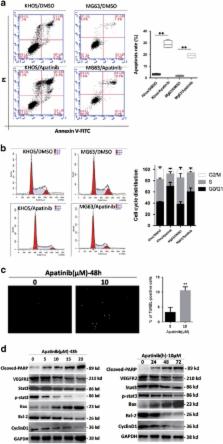
          The cure rate of osteosarcoma has not improved in the past 30 years. The search for new treatments and drugs is urgently needed. Apatinib is a high selectivity inhibitor of vascular endothelial growth factor receptor-2 (VEGFR2) tyrosine kinase, exerting promising antitumoral effect in various tumors. The antitumor effect of Apatinib in human osteosarcoma has never been reported. We investigated the effects of Apatinib in osteosarcoma in vitro and in vivo. Osteosarcoma patients with high levels of VEGFR2 have poor prognosis. Apatinib can inhibit cell growth of osteosarcoma cells. In addition to cycle arrest and apoptosis, Apatinib induces autophagy. Interestingly, inhibition of autophagy increased Apatinib-induced apoptosis in osteosarcoma cells. Immunoprecipitation confirmed direct binding between VEGFR2 and signal transducer and activator of transcription 3 (STAT3). Downregulation of VEGFR2 by siRNA resulted in STAT3 inhibition in KHOS cells. VEGFR2 and STAT3 are inhibited by Apatinib in KHOS cells, and STAT3 act downstream of VEGFR2. STAT3 and BCL-2 were downregulated by Apatinib. STAT3 knockdown by siRNA reinforced autophagy and apoptosis induced by Apatinib. BCL-2 inhibits autophagy and was apoptosis restrained by Apatinib too. Overexpression of BCL-2 decreased Apatinib-induced apoptosis and autophagy. Apatinib repressed the expression of STAT3 and BCL-2 and suppressed the growth of osteosarcoma in vivo. To sum up, deactivation of VEGFR2/STAT3/BCL-2 signal pathway leads to Apatinib-induced growth inhibition of osteosarcoma.

          Related collections

          Most cited references33

          • Record: found
          • Abstract: found
          • Article: not found

          Role of the vascular endothelial growth factor pathway in tumor growth and angiogenesis.

          New blood vessel formation (angiogenesis) is a fundamental event in the process of tumor growth and metastatic dissemination. Hence, the molecular basis of tumor angiogenesis has been of keen interest in the field of cancer research. The vascular endothelial growth factor (VEGF) pathway is well established as one of the key regulators of this process. The VEGF/VEGF-receptor axis is composed of multiple ligands and receptors with overlapping and distinct ligand-receptor binding specificities, cell-type expression, and function. Activation of the VEGF-receptor pathway triggers a network of signaling processes that promote endothelial cell growth, migration, and survival from pre-existing vasculature. In addition, VEGF mediates vessel permeability, and has been associated with malignant effusions. More recently, an important role for VEGF has emerged in mobilization of endothelial progenitor cells from the bone marrow to distant sites of neovascularization. The well-established role of VEGF in promoting tumor angiogenesis and the pathogenesis of human cancers has led to the rational design and development of agents that selectively target this pathway. Studies with various anti-VEGF/VEGF-receptor therapies have shown that these agents can potently inhibit angiogenesis and tumor growth in preclinical models. Recently, an anti-VEGF antibody (bevacizumab), when used in combination with chemotherapy, was shown to significantly improve survival and response rates in patients with metastatic colorectal cancer and thus, validate VEGF pathway inhibitors as an important new treatment modality in cancer therapy.
            Bookmark
            • Record: found
            • Abstract: found
            • Article: not found

            Life and death partners: apoptosis, autophagy and the cross-talk between them.

            It is not surprising that the demise of a cell is a complex well-controlled process. Apoptosis, the first genetically programmed death process identified, has been extensively studied and its contribution to the pathogenesis of disease well documented. Yet, apoptosis does not function alone to determine a cell's fate. More recently, autophagy, a process in which de novo-formed membrane-enclosed vesicles engulf and consume cellular components, has been shown to engage in a complex interplay with apoptosis. In some cellular settings, it can serve as a cell survival pathway, suppressing apoptosis, and in others, it can lead to death itself, either in collaboration with apoptosis or as a back-up mechanism when the former is defective. The molecular regulators of both pathways are inter-connected; numerous death stimuli are capable of activating either pathway, and both pathways share several genes that are critical for their respective execution. The cross-talk between apoptosis and autophagy is therefore quite complex, and sometimes contradictory, but surely critical to the overall fate of the cell. Furthermore, the cross-talk is a key factor in the outcome of death-related pathologies such as cancer, its development and treatment.
              Bookmark
              • Record: found
              • Abstract: found
              • Article: found
              Is Open Access

              Dissociation of Bcl-2–Beclin1 Complex by Activated AMPK Enhances Cardiac Autophagy and Protects Against Cardiomyocyte Apoptosis in Diabetes

              Diabetic cardiomyopathy is associated with suppression of cardiac autophagy, and activation of AMP-activated protein kinase (AMPK) restores cardiac autophagy and prevents cardiomyopathy in diabetic mice, albeit by an unknown mechanism. We hypothesized that AMPK-induced autophagy ameliorates diabetic cardiomyopathy by inhibiting cardiomyocyte apoptosis and examined the effects of AMPK on the interaction between Beclin1 and Bcl-2, a switch between autophagy and apoptosis, in diabetic mice and high glucose–treated H9c2 cardiac myoblast cells. Exposure of H9c2 cells to high glucose reduced AMPK activity, inhibited Jun NH2-terminal kinase 1 (JNK1)–B-cell lymphoma 2 (Bcl-2) signaling, and promoted Beclin1 binding to Bcl-2. Conversely, activation of AMPK by metformin stimulated JNK1–Bcl-2 signaling and disrupted the Beclin1–Bcl-2 complex. Activation of AMPK, which normalized cardiac autophagy, attenuated high glucose–induced apoptosis in cultured H9c2 cells. This effect was attenuated by inhibition of autophagy. Finally, chronic administration of metformin in diabetic mice restored cardiac autophagy by activating JNK1–Bcl-2 pathways and dissociating Beclin1 and Bcl-2. The induction of autophagy protected against cardiac apoptosis and improved cardiac structure and function in diabetic mice. We concluded that dissociation of Bcl-2 from Beclin1 may be an important mechanism for preventing diabetic cardiomyopathy via AMPK activation that restores autophagy and protects against cardiac apoptosis.
                Bookmark

                Author and article information

                Journal
                Cell Death Dis
                Cell Death Dis
                Cell Death & Disease
                Nature Publishing Group
                2041-4889
                August 2017
                24 August 2017
                1 August 2017
                : 8
                : 8
                : e3015
                Affiliations
                [1 ]Musculoskeletal Tumor Center, Peking University People’s Hospital , Beijing, People’s Republic of China
                [2 ]Beijing Key Laboratory of Musculoskeletal Tumor , Beijing, People’s Republic of China
                [3 ]Department of Pathology, Peking University People’s Hospital , Beijing, People’s Republic of China
                Author notes
                [* ]Musculoskeletal Tumor Center, Peking University People’s Hospital , No.11 Xizhimen South Street, Beijing 100044, People’s Republic of China. Tel: +86 10 883 244 71; Fax: +86 10 883 785 44; E-mail: bonetumor@ 123456163.com
                Article
                cddis2017422
                10.1038/cddis.2017.422
                5596600
                28837148
                c35b2291-404a-46fb-9770-d9cec74ec8ae
                Copyright © 2017 The Author(s)

                Cell Death and Disease is an open-access journal published by Nature Publishing Group. This work is licensed under a Creative Commons Attribution 4.0 International License. The images or other third party material in this article are included in the article’s Creative Commons license, unless indicated otherwise in the credit line; if the material is not included under the Creative Commons license, users will need to obtain permission from the license holder to reproduce the material. To view a copy of this license, visit http://creativecommons.org/licenses/by/4.0/

                History
                : 23 May 2017
                : 20 July 2017
                : 24 July 2017
                Categories
                Original Article

                Cell biology
                Cell biology

                Comments

                Comment on this article