Inviting an author to review:
Find an author and click ‘Invite to review selected article’ near their name.
Search for authorsSearch for similar articles
6
views
0
recommends
+1 Recommend
0 collections
    0
    shares
      • Record: found
      • Abstract: found
      • Article: found
      Is Open Access

      microRNAs in the Regulation of Melanogenesis

      review-article

      Read this article at

      Bookmark
          There is no author summary for this article yet. Authors can add summaries to their articles on ScienceOpen to make them more accessible to a non-specialist audience.

          Abstract

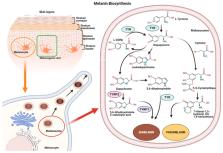
          Melanogenesis is the process leading to the synthesis of melanin, the main substance that influences skin color and plays a pivotal role against UV damage. Altered melanogenesis is observed in several pigmentation disorders. Melanogenesis occurs in specialized cells called melanocytes, physically and functionally related by means of autocrine and paracrine interplay to other skin cell types. Several external and internal factors control melanin biosynthesis and operate through different intracellular signaling pathways, which finally leads to the regulation of microphthalmia-associated transcription factor ( MITF), the key transcription factor involved in melanogenesis and the expression of the main melanogenic enzymes, including TYR, TYRP-1, and TYRP-2. Epigenetic factors, including microRNAs (miRNAs), are involved in melanogenesis regulation. miRNAs are small, single-stranded, non-coding RNAs, of approximately 22 nucleotides in length, which control cell behavior by regulating gene expression, mainly by binding the 3′ untranslated region (3′-UTR) of target mRNAs. This review collects data on the miRNAs involved in melanogenesis and how these miRNAs can modulate target gene expression. Bringing to light the biological function of miRNAs could lead to a wider understanding of epigenetic melanogenesis regulation and its dysregulation. This knowledge may constitute the basis for developing innovative treatment approaches for pigmentation dysregulation.

          Related collections

          Most cited references100

          • Record: found
          • Abstract: found
          • Article: not found

          Exosome-mediated transfer of mRNAs and microRNAs is a novel mechanism of genetic exchange between cells.

          Exosomes are vesicles of endocytic origin released by many cells. These vesicles can mediate communication between cells, facilitating processes such as antigen presentation. Here, we show that exosomes from a mouse and a human mast cell line (MC/9 and HMC-1, respectively), as well as primary bone marrow-derived mouse mast cells, contain RNA. Microarray assessments revealed the presence of mRNA from approximately 1300 genes, many of which are not present in the cytoplasm of the donor cell. In vitro translation proved that the exosome mRNAs were functional. Quality control RNA analysis of total RNA derived from exosomes also revealed presence of small RNAs, including microRNAs. The RNA from mast cell exosomes is transferable to other mouse and human mast cells. After transfer of mouse exosomal RNA to human mast cells, new mouse proteins were found in the recipient cells, indicating that transferred exosomal mRNA can be translated after entering another cell. In summary, we show that exosomes contain both mRNA and microRNA, which can be delivered to another cell, and can be functional in this new location. We propose that this RNA is called "exosomal shuttle RNA" (esRNA).
            Bookmark
            • Record: found
            • Abstract: found
            • Article: found
            Is Open Access

            Overview of MicroRNA Biogenesis, Mechanisms of Actions, and Circulation

            MicroRNAs (miRNAs) are a class of non-coding RNAs that play important roles in regulating gene expression. The majority of miRNAs are transcribed from DNA sequences into primary miRNAs and processed into precursor miRNAs, and finally mature miRNAs. In most cases, miRNAs interact with the 3′ untranslated region (3′ UTR) of target mRNAs to induce mRNA degradation and translational repression. However, interaction of miRNAs with other regions, including the 5′ UTR, coding sequence, and gene promoters, have also been reported. Under certain conditions, miRNAs can also activate translation or regulate transcription. The interaction of miRNAs with their target genes is dynamic and dependent on many factors, such as subcellular location of miRNAs, the abundancy of miRNAs and target mRNAs, and the affinity of miRNA-mRNA interactions. miRNAs can be secreted into extracellular fluids and transported to target cells via vesicles, such as exosomes, or by binding to proteins, including Argonautes. Extracellular miRNAs function as chemical messengers to mediate cell-cell communication. In this review, we provide an update on canonical and non-canonical miRNA biogenesis pathways and various mechanisms underlying miRNA-mediated gene regulations. We also summarize the current knowledge of the dynamics of miRNA action and of the secretion, transfer, and uptake of extracellular miRNAs.
              Bookmark
              • Record: found
              • Abstract: not found
              • Article: not found

              The C. elegans heterochronic gene lin-4 encodes small RNAs with antisense complementarity to lin-14

                Bookmark

                Author and article information

                Contributors
                Role: Academic Editor
                Journal
                Int J Mol Sci
                Int J Mol Sci
                ijms
                International Journal of Molecular Sciences
                MDPI
                1422-0067
                05 June 2021
                June 2021
                : 22
                : 11
                : 6104
                Affiliations
                [1 ]Morgan S.r.l, R&D, 36050 Monteviale, VI, Italy; yekatsiaryna.hushcha@ 123456gmail.com or
                [2 ]Department of Medical Sciences, Section of Experimental Medicine, School of Medicine, University of Ferrara, 64b, Fossato di Mortara Street, 44121 Ferrara, Italy; irene01.blo@ 123456edu.unife.it (I.B.); lucia.otongonzalez@ 123456unife.it (L.O.-G.); dmrgli@ 123456unife.it (G.D.M.); fernanda.martini@ 123456unife.it (F.M.); mauro.tognon@ 123456unife.it (M.T.)
                [3 ]Laboratory for Technologies of Advanced Therapies (LTTA), University of Ferrara, 44121 Ferrara, Italy
                Author notes
                [* ]Correspondence: monica.demattei@ 123456unife.it ; Tel.: +39-0532-455534
                Author information
                https://orcid.org/0000-0002-8515-6754
                https://orcid.org/0000-0001-6739-6423
                https://orcid.org/0000-0002-5789-0601
                Article
                ijms-22-06104
                10.3390/ijms22116104
                8201055
                34198907
                c80fe5b4-56b1-4fce-95ec-993d0ce901db
                © 2021 by the authors.

                Licensee MDPI, Basel, Switzerland. This article is an open access article distributed under the terms and conditions of the Creative Commons Attribution (CC BY) license ( https://creativecommons.org/licenses/by/4.0/).

                History
                : 16 April 2021
                : 03 June 2021
                Categories
                Review

                Molecular biology
                melanocyte,melanogenesis,microrna,skin pigmentation
                Molecular biology
                melanocyte, melanogenesis, microrna, skin pigmentation

                Comments

                Comment on this article