3
views
0
recommends
+1 Recommend
0 collections
    0
    shares
      • Record: found
      • Abstract: found
      • Article: found
      Is Open Access

      Comparison of paired cerebrospinal fluid and serum cell‐free mitochondrial and nuclear DNA with copy number and fragment length

      research-article

      Read this article at

      Bookmark
          There is no author summary for this article yet. Authors can add summaries to their articles on ScienceOpen to make them more accessible to a non-specialist audience.

          Abstract

          Background

          Most studies on cell‐free DNA (cfDNA) were only for single body fluids; however, the differences in cfDNA distribution between two body fluids are rarely reported. Hence, in this work, we compared the differences in cfDNA distribution between cerebrospinal fluid (CSF) and serum of patients with brain‐related diseases.

          Methods

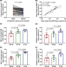
          The fragment length of cfDNA was determined by using Agilent 2100 Bioanalyzer. The copy numbers of cell‐free mitochondrial DNA (cf‐mtDNA) and cell‐free nuclear DNA (cf‐nDNA) were determined by using real‐time quantitative PCR (qPCR) and droplet digital PCR (ddPCR) with three pairs of mitochondrial ND1 and nuclear GAPDH primers, respectively.

          Results

          There were short (~60 bp), medium (~167 bp), and long (>250 bp) cfDNA fragment length distributions totally obtained from CSF and serum using Agilent 2100 Bioanalyzer. The results of both qPCR and ddPCR confirmed the existence of these three cfDNA fragment ranges in CSF and serum. According to qPCR, the copy numbers of long cf‐mtDNA, medium, and long cf‐nDNA in CSF were significantly higher than in paired serum. In CSF, only long cf‐mtDNA's copy numbers were higher than long cf‐nDNA. But in serum, the copy numbers of medium and long cf‐mtDNA were higher than the corresponding cf‐nDNA.

          Conclusion

          The cf‐nDNA and cf‐mtDNA with different fragment lengths differentially distributed in the CSF and serum of patients with brain disorders, which might serve as a biomarker of human brain diseases.

          Related collections

          Most cited references36

          • Record: found
          • Abstract: found
          • Article: found
          Is Open Access

          Origins, structures, and functions of circulating DNA in oncology

          While various clinical applications especially in oncology are now in progress such as diagnosis, prognosis, therapy monitoring, or patient follow-up, the determination of structural characteristics of cell-free circulating DNA (cirDNA) are still being researched. Nevertheless, some specific structures have been identified and cirDNA has been shown to be composed of many “kinds.” This structural description goes hand-in-hand with the mechanisms of its origins such as apoptosis, necrosis, active release, phagocytosis, and exocytose. There are multiple structural forms of cirDNA depending upon the mechanism of release: particulate structures (exosomes, microparticles, apoptotic bodies) or macromolecular structures (nucleosomes, virtosomes/proteolipidonucleic acid complexes, DNA traps, links with serum proteins or to the cell-free membrane parts). In addition, cirDNA concerns both nuclear and/or mitochondrial DNA with both species exhibiting different structural characteristics that potentially reveal different forms of biological stability or diagnostic significance. This review focuses on the origins, structures and functional aspects that are paradoxically less well described in the literature while numerous reviews are directed to the clinical application of cirDNA. Differentiation of the various structures and better knowledge of the fate of cirDNA would considerably expand the diagnostic power of cirDNA analysis especially with regard to the patient follow-up enlarging the scope of personalized medicine. A better understanding of the subsequent fate of cirDNA would also help in deciphering its functional aspects such as their capacity for either genometastasis or their pro-inflammatory and immunological effects.
            Bookmark
            • Record: found
            • Abstract: found
            • Article: not found

            Predominant hematopoietic origin of cell-free DNA in plasma and serum after sex-mismatched bone marrow transplantation.

            Despite current interest in the biology and diagnostic applications of cell-free DNA in plasma and serum, the cellular origin of this DNA is poorly understood. We used a sex-mismatched bone marrow transplantation model to study the relative contribution of hematopoietic and nonhematopoietic cells to circulating DNA. We studied 22 sex-mismatched bone marrow transplantation patients. Paired buffy coat and plasma samples were obtained from all 22 patients. Matching serum samples were also obtained from seven of them. Plasma DNA, serum DNA, and buffy coat were quantified by real-time PCR of the SRY and beta-globin gene DNA. To investigate the effects of blood drawing and other preanalytical variables on plasma DNA concentrations, blood samples were also collected from 14 individuals who had not received transplants. The effects of blood sampling by syringe and needle, centrifugation, and time delay in blood processing were studied. The median percentage of Y-chromosome DNA in the plasma in female patients receiving bone marrow from male donors (59.5%) differed significantly (P <0.001) from that in the male patients receiving bone marrow from female donors (6.9%). This indicated that plasma DNA in the bone marrow transplantation recipients was predominantly of donor origin. Compared with paired plasma samples, serum samples had a median 14-fold higher DNA concentration, with the additional DNA being of donor origin. Control experiments indicated that none of the three tested preanalytical variables contributed to a significant change in cell-free DNA concentration. After bone marrow transplantation, the DNA in plasma and serum is predominantly hematopoietic in origin. Apart from the biological implications of this observation, this finding suggests that plasma and serum can be used as alternative materials for the study of postbone marrow transplantation chimerism.
              Bookmark
              • Record: found
              • Abstract: found
              • Article: not found

              Cell-free DNA in human blood plasma: length measurements in patients with pancreatic cancer and healthy controls.

              The amount of non-cell-associated DNA free in blood plasma from pancreatic cancer patients usually exceeds that from healthy donors. We have evaluated the plasma DNA by gel electrophoresis and measured the variation in length of soluble DNA fragments by electron microscopy in plasma from three patients with pancreatic cancer and from three healthy controls. Whereas electrophoresis of nick-translated DNA isolated from plasma obtained from healthy controls showed autoradiographic bands at sizes equivalent to whole-number multiples (1-5x) of nucleosomal DNA (185-200 bp), in the samples obtained from pancreatic cancer patients, stronger ladder patterns appeared. Likewise, strand length distributions of DNA (DNA-SL) in the two groups differ. The DNA-SL distribution data include 2,752 measurements made from cancer patient plasma and 3,291 for control plasma. The shortest DNA-SL measured approximately 30 nm (approximately 88 bp calculated at 0.34 nm/bp) and the largest approximately 28,000 nm (>80,000 bp), with 50% of all lengths measuring between 100 and 900 nm long. The average plasma DNA-SL in controls (311 nm; median, 273 nm) exceeded that in cancer patients (231 nm; median, 185 nm). Small excesses of DNA at approximately 63, approximately 126, approximately 189, approximately 252, and approximately 315 nm, corresponding to small multiples of lengths associated with nucleosomes, were more prominent in the cancer patient plasma than in the healthy control plasma. This study provides evidence indicating differences in non-cell-associated DNA in plasma between cancer patients and healthy controls and indicates that a significant amount of this DNA is probably derived from apoptosis in neoplastic and/or normal cells.
                Bookmark

                Author and article information

                Contributors
                jxlu313@163.com
                wuwenhe2000@163.com , wwh2000@wmu.edu.cn
                Journal
                J Clin Lab Anal
                J. Clin. Lab. Anal
                10.1002/(ISSN)1098-2825
                JCLA
                Journal of Clinical Laboratory Analysis
                John Wiley and Sons Inc. (Hoboken )
                0887-8013
                1098-2825
                13 February 2020
                June 2020
                : 34
                : 6 ( doiID: 10.1002/jcla.v34.6 )
                : e23238
                Affiliations
                [ 1 ] Key Laboratory of Laboratory Medicine Ministry of Education Zhejiang Provincial Key Laboratory of Medical Genetics School of Laboratory Medicine and Life Sciences Wenzhou Medical University Wenzhou China
                [ 2 ] Department of Clinical Laboratory Wenzhou People's Hospital Wenzhou China
                [ 3 ] Hangzhou Medical College Hangzhou China
                Author notes
                [*] [* ] Correspondence

                Jianxin Lyu and Wenhe Wu, Key Laboratory of Laboratory Medicine, Ministry of Education, Zhejiang Provincial Key Laboratory of Medical Genetics, School of Laboratory Medicine and Life Sciences, Wenzhou Medical University, Wenzhou, Zhejiang 325035, China.

                Emails: jxlu313@ 123456163.com (JL); wuwenhe2000@ 123456163.com and wwh2000@ 123456wmu.edu.cn (WW)

                Author information
                https://orcid.org/0000-0002-9114-5111
                Article
                JCLA23238
                10.1002/jcla.23238
                7307366
                32052892
                cbeae7f9-2f1b-4a3f-b1da-265c89bfcc5e
                © 2020 The Authors. Journal of Clinical Laboratory Analysis published by Wiley Periodicals, Inc.

                This is an open access article under the terms of the http://creativecommons.org/licenses/by/4.0/ License, which permits use, distribution and reproduction in any medium, provided the original work is properly cited.

                History
                : 11 August 2019
                : 11 January 2020
                : 21 January 2020
                Page count
                Figures: 6, Tables: 3, Pages: 12, Words: 7280
                Funding
                Funded by: Zhejiang Xinmiao Talent Projects
                Award ID: 2017R413073
                Funded by: Wenzhou Science and Technology Planning Project
                Award ID: Y20170249
                Funded by: National Natural Science Foundation of China , open-funder-registry 10.13039/501100001809;
                Award ID: 81101148
                Award ID: 81670712
                Categories
                Research Article
                Research Articles
                Custom metadata
                2.0
                June 2020
                Converter:WILEY_ML3GV2_TO_JATSPMC version:5.8.4 mode:remove_FC converted:22.06.2020

                Clinical chemistry
                cell‐free mitochondrial dna,cell‐free nuclear dna,cerebrospinal fluid,copy number,fragment length,serum

                Comments

                Comment on this article