Inviting an author to review:
Find an author and click ‘Invite to review selected article’ near their name.
Search for authorsSearch for similar articles
51
views
0
recommends
+1 Recommend
0 collections
    0
    shares
      • Record: found
      • Abstract: found
      • Article: not found

      Microfluidic Control of Cell Pairing and Fusion

      research-article

      Read this article at

      ScienceOpenPublisherPMC
      Bookmark
          There is no author summary for this article yet. Authors can add summaries to their articles on ScienceOpen to make them more accessible to a non-specialist audience.

          Abstract

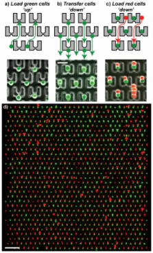
          Cell fusion has been used for many different purposes, including generation of hybridomas and reprogramming of somatic cells. The fusion step represents the key event in initiation of these procedures. Standard fusion techniques, however, provide poor and random cell contact, leading to low yields. We present here a microfluidic device to trap and properly pair thousands of cells. Using this device we were able to pair different cell types, including fibroblasts, mouse embryonic stem cells (mESCs), and myeloma cells, achieving pairing efficiencies up to 70%. The device is compatible with both chemical and electrical fusion protocols. We observed that electrical fusion was more efficient than chemical fusion, with membrane reorganization efficiencies of up to 89%. We achieved greater than 50% properly paired and fused cells over the entire device, 5× greater than a commercial electrofusion chamber, and were able to observe reprogramming in hybrids between mESCs and mouse embryonic fibroblasts.

          Related collections

          Most cited references29

          • Record: found
          • Abstract: found
          • Article: not found

          Viable offspring derived from fetal and adult mammalian cells.

          Fertilization of mammalian eggs is followed by successive cell divisions and progressive differentiation, first into the early embryo and subsequently into all of the cell types that make up the adult animal. Transfer of a single nucleus at a specific stage of development, to an enucleated unfertilized egg, provided an opportunity to investigate whether cellular differentiation to that stage involved irreversible genetic modification. The first offspring to develop from a differentiated cell were born after nuclear transfer from an embryo-derived cell line that had been induced to become quiescent. Using the same procedure, we now report the birth of live lambs from three new cell populations established from adult mammary gland, fetus and embryo. The fact that a lamb was derived from an adult cell confirms that differentiation of that cell did not involve the irreversible modification of genetic material required for development to term. The birth of lambs from differentiated fetal and adult cells also reinforces previous speculation that by inducing donor cells to become quiescent it will be possible to obtain normal development from a wide variety of differentiated cells.
            Bookmark
            • Record: found
            • Abstract: found
            • Article: not found

            In vitro reprogramming of fibroblasts into a pluripotent ES-cell-like state.

            Nuclear transplantation can reprogramme a somatic genome back into an embryonic epigenetic state, and the reprogrammed nucleus can create a cloned animal or produce pluripotent embryonic stem cells. One potential use of the nuclear cloning approach is the derivation of 'customized' embryonic stem (ES) cells for patient-specific cell treatment, but technical and ethical considerations impede the therapeutic application of this technology. Reprogramming of fibroblasts to a pluripotent state can be induced in vitro through ectopic expression of the four transcription factors Oct4 (also called Oct3/4 or Pou5f1), Sox2, c-Myc and Klf4. Here we show that DNA methylation, gene expression and chromatin state of such induced reprogrammed stem cells are similar to those of ES cells. Notably, the cells-derived from mouse fibroblasts-can form viable chimaeras, can contribute to the germ line and can generate live late-term embryos when injected into tetraploid blastocysts. Our results show that the biological potency and epigenetic state of in-vitro-reprogrammed induced pluripotent stem cells are indistinguishable from those of ES cells.
              Bookmark
              • Record: found
              • Abstract: found
              • Article: not found

              Plasticity of the differentiated state.

              Heterokaryons provide a model system in which to examine how tissue-specific phenotypes arise and are maintained. When muscle cells are fused with nonmuscle cells, muscle gene expression is activated in the nonmuscle cell type. Gene expression was studied either at a single cell level with monoclonal antibodies or in mass cultures at a biochemical and molecular level. In all of the nonmuscle cell types tested, including representatives of different embryonic lineages, phenotypes, and developmental stages, muscle gene expression was induced. Differences among cell types in the kinetics, frequency, and gene dosage requirements for gene expression provide clues to the underlying regulatory mechanisms. These results show that the expression of genes in the nuclei of differentiated cells is remarkably plastic and susceptible to modulation by the cytoplasm. The isolation of the genes encoding the tissue-specific trans-acting regulators responsible for muscle gene activation should now be possible.
                Bookmark

                Author and article information

                Journal
                101215604
                32338
                Nat Methods
                Nature methods
                1548-7091
                1548-7105
                12 December 2011
                4 January 2009
                February 2009
                4 January 2012
                : 6
                : 2
                : 147-152
                Affiliations
                [1 ]Research Laboratory of Electronics, 50 Vassar Street, Massachusetts Institute of Technology, Cambridge, MA 02139
                [2 ]Microsystems Technology Laboratory, 60 Vassar Street, Massachusetts Institute of Technology, Cambridge, MA 02139
                [3 ]Whitehead Institute for Biomedical Research, Nine Cambridge Center, Cambridge, MA 02142
                [4 ]Department of Biology, Massachusetts Institute of Technology, 77 Massachusetts Avenue, Cambridge, Massachusetts 02139
                [5 ]Electrical Engineering and Computer Science Department, 77 Massachusetts Avenue, Massachusetts Institute of Technology, Cambridge, MA 02139
                Author notes
                Corresponding Authors: Joel Voldman, voldman@ 123456mit.edu , Department of Electrical Engineering & Computer Science, Massachusetts Institute of Technology, Room 36-824, 77 Massachusetts Ave, Cambridge, MA 02139, Ph: 617.253.2094, Fx: 617.258.5846. Rudolf Jaenisch, jaenisch@ 123456wi.mit.edu , Member, Whitehead Institute for Biomedical Research, Professor of Biology, Massachusetts Institute of Technology, Nine Cambridge Center, Cambridge, Massachusetts 02147-1479, Ph: 617.258.5186, Fx: 617.258.6505
                [¶]

                These authors contributed equally to this work.

                Article
                nihpa80614
                10.1038/nmeth.1290
                3251011
                19122668
                cf2019b8-f826-4c25-b2ff-13791434c33e
                History
                Funding
                Funded by: National Cancer Institute : NCI
                Award ID: R01 CA087869-08 || CA
                Categories
                Article

                Life sciences
                Life sciences

                Comments

                Comment on this article