23
views
0
recommends
+1 Recommend
0 collections
    0
    shares
      • Record: found
      • Abstract: found
      • Article: found
      Is Open Access

      Phosphorylated SATB1 is associated with the progression and prognosis of glioma

      research-article

      Read this article at

      Bookmark
          There is no author summary for this article yet. Authors can add summaries to their articles on ScienceOpen to make them more accessible to a non-specialist audience.

          Abstract

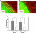
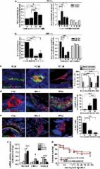
          Special AT-rich sequence-binding protein 1 (SATB1) is a global chromatin organizer and gene regulator, and high expression of SATB1 is associated with progression and poor prognosis in several malignancies. Here, we examine the expression pattern of SATB1 in glioma. Microarray analysis of 127 clinical samples showed that SATB1 mRNA was expressed at lower levels in highly malignant glioblastoma multiforme (GBM) than in low-grade glioma and normal brain tissue. This result was further confirmed by real-time RT-PCR in the clinical samples, three GBM cell lines, primary SU3 glioma cells and tumor cells harvested by laser-capture microdissection. Consistent with the mRNA levels, SATB1 protein expression was downregulated in high-grade glioma, as shown by western blotting. However, phospho-SATB1 levels showed an opposite pattern, with a significant increase in these tumors. Immunohistochemical analysis of phospho-SATB1 expression in tissue microarrays with tumors from 122 glioma cases showed that phospho-SATB1 expression was significantly associated with high histological grade and poor survival by Kaplan–Meier analysis. In vitro transfection analysis showed that phospho-SATB1 DNA binding has a key role in regulating the proliferation and invasion of glioma cells. The effect of SATB1 in glioma cell is mainly histone deacetylase (HDAC1)-dependent. We conclude that phospho-SATB1, but not SATB1 mRNA expression, is associated with the progression and prognosis of glioma. By interaction with HDAC1, phospho-SATB1 contributes to the invasive and proliferative phenotype of GBM cells.

          Related collections

          Most cited references26

          • Record: found
          • Abstract: found
          • Article: not found

          Inactivation of the DNA repair gene O6-methylguanine-DNA methyltransferase by promoter hypermethylation is a common event in primary human neoplasia.

          The DNA repair protein O6-methylguanine DNA methyltransferase (MGMT) removes alkyl adducts from the O6 position of guanine. MGMT expression is decreased in some tumor tissues, and lack of activity has been observed in some cell lines. Loss of expression is rarely due to deletion, mutation, or rearrangement of the MGMT gene, but methylation of discrete regions of the CpG island of MGMT has been associated with the silencing of the gene in cell lines. We used methylation-specific PCR to study the promoter methylation of the MGMT gene. All normal tissues and expressing cancer cell lines were unmethylated, whereas nonexpressing cancer cell lines were methylated. Among the more than 500 primary human tumors examined, MGMT hypermethylation was present in a subset of specific types of cancer. In gliomas and colorectal carcinomas, aberrant methylation was detected in 40% of the tumors, whereas in non-small cell lung carcinomas, lymphomas, and head and neck carcinomas, this alteration was found in 25% of the tumors. MGMT methylation was found rarely or not at all in other tumor types. We also analyzed MGMT expression by immunohistochemistry in relation to the methylation status in 31 primary tumors. The presence of aberrant hypermethylation was associated with loss of MGMT protein, in contrast to retention of protein in the majority of tumors without aberrant hypermethylation. Our results suggest that epigenetic inactivation of MGMT plays an important role in primary human neoplasia.
            Bookmark
            • Record: found
            • Abstract: found
            • Article: found
            Is Open Access

            Astrocytes Enhance the Invasion Potential of Glioblastoma Stem-Like Cells

            Glioblastomas (GBMs) are characterized as highly invasive; the contribution of GBM stem-like cells (GSCs) to the invasive phenotype, however, has not been completely defined. Towards this end, we have defined the invasion potential of CD133+ GSCs and their differentiated CD133− counterparts grown under standard in vitro conditions and in co-culture with astrocytes. Using a trans-well assay, astrocytes or astrocyte conditioned media in the bottom chamber significantly increased the invasion of GSCs yet had no effect on CD133− cells. In addition, a monolayer invasion assay showed that the GSCs invaded farther into an astrocyte monolayer than their differentiated progeny. Gene expression profiles were generated from two GSC lines grown in trans-well culture with astrocytes in the bottom chamber or directly in contact with astrocyte monolayers. In each co-culture model, genes whose expression was commonly increased in both GSC lines involved cell movement and included a number of genes that have been previously associated with tumor cell invasion. Similar gene expression modifications were not detected in CD133− cells co-cultured under the same conditions with astrocytes. Finally, evaluation of the secretome of astrocytes grown in monolayer identified a number of chemokines and cytokines associated with tumor cell invasion. These data suggest that astrocytes enhance the invasion of CD133+ GSCs and provide additional support for a critical role of brain microenvironment in the regulation of GBM biology.
              Bookmark
              • Record: found
              • Abstract: found
              • Article: found
              Is Open Access

              Wnt activation promotes neuronal differentiation of Glioblastoma

              One of the biggest challenges in tumour research is the possibility to reprogram cancer cells towards less aggressive phenotypes. In this study, we reprogrammed primary Glioblastoma multiforme (GBM)-derived cells towards a more differentiated and less oncogenic phenotype by activating the Wnt pathway in a hypoxic microenvironment. Hypoxia usually correlates with malignant behaviours in cancer cells, but it has been recently involved, together with Wnt signalling, in the differentiation of embryonic and neural stem cells. Here, we demonstrate that treatment with Wnt ligands, or overexpression of β-catenin, mediate neuronal differentiation and halt proliferation in primary GBM cells. An hypoxic environment cooperates with Wnt-induced differentiation, in line with our finding that hypoxia inducible factor-1α (HIF-1α) is instrumental and required to sustain the expression of β-catenin transcriptional partners TCF-1 and LEF-1. In addition, we also found that Wnt-induced GBM cell differentiation inhibits Notch signalling, and thus gain of Wnt and loss of Notch cooperate in the activation of a pro-neuronal differentiation program. Intriguingly, the GBM sub-population enriched of cancer stem cells (CD133+ fraction) is the primary target of the pro-differentiating effects mediated by the crosstalk between HIF-1α, Wnt, and Notch signalling. By using zebrafish transgenics and mutants as model systems to visualize and manipulate in vivo the Wnt pathway, we confirm that Wnt pathway activation is able to promote neuronal differentiation and inhibit Notch signalling of primary human GBM cells also in this in vivo set-up. In conclusion, these findings shed light on an unsuspected crosstalk between hypoxia, Wnt and Notch signalling in GBM, and suggest the potential to manipulate these microenvironmental signals to blunt GBM malignancy.
                Bookmark

                Author and article information

                Journal
                Cell Death Dis
                Cell Death Dis
                Cell Death & Disease
                Nature Publishing Group
                2041-4889
                October 2013
                31 October 2013
                1 October 2013
                : 4
                : 10
                : e901
                Affiliations
                [1 ]Department of Neurosurgery, The First Hospital of China Medical University , Shenyang, China
                Author notes
                [* ]Department of Neurosurgery, The First Hospital of China Medical University , Nanjing Street 155, Heping District, Shenyang 110001, China. Tel: +86 024 83283129; Fax: +86 024 83283133; E-mail: cmuwuanhua@ 123456yahoo.com.cn
                [2]

                These authors contribute equally to this work.

                Article
                cddis2013433
                10.1038/cddis.2013.433
                3920943
                24176859
                d06d1c51-5de8-4197-a1c7-8871266426cb
                Copyright © 2013 Macmillan Publishers Limited

                This work is licensed under a Creative Commons Attribution-NonCommercial-NoDerivs 3.0 Unported License. To view a copy of this license, visit http://creativecommons.org/licenses/by-nc-nd/3.0/

                History
                : 18 July 2013
                : 17 September 2013
                : 02 October 2013
                Categories
                Original Article

                Cell biology
                glioblastoma multiforme,special at-rich sequence-binding protein 1,phosphorylation,prognosis

                Comments

                Comment on this article