24
views
0
recommends
+1 Recommend
0 collections
    0
    shares
      • Record: found
      • Abstract: found
      • Article: not found

      Phosphorylation at S365 is a gatekeeper event that changes the structure of Cx43 and prevents down-regulation by PKC

      research-article

      Read this article at

      ScienceOpenPublisherPMC
      Bookmark
          There is no author summary for this article yet. Authors can add summaries to their articles on ScienceOpen to make them more accessible to a non-specialist audience.

          Abstract

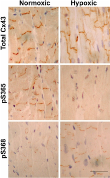
          Phosphorylation at unspecified sites is known to regulate the life cycle (assembly, gating, and turnover) of the gap junction protein, Cx43. In this paper, we show that Cx43 is phosphorylated on S365 in cultured cells and heart tissue. Nuclear magnetic resonance structural studies of the C-terminal region of Cx43 with an S365D mutation indicate that it forms a different stable conformation than unphosphorylated wild-type Cx43. Immunolabeling with an antibody specific for Cx43 phosphorylated at S365 shows staining on gap junction structures in heart tissue that is lost upon hypoxia when Cx43 is no longer specifically localized to the intercalated disk. Efficient phosphorylation at S368, an important Cx43 channel regulatory event that increases during ischemia or PKC activation, depends on S365 being unphosphorylated. Thus, phosphorylation at S365 can serve a “gatekeeper” function that may represent a mechanism to protect cells from ischemia and phorbol ester-induced down-regulation of channel conductance.

          Related collections

          Most cited references38

          • Record: found
          • Abstract: found
          • Article: not found

          RADIOAUTOGRAPHIC STUDIES OF CHOLINE INCORPORATION INTO PERIPHERAL NERVE MYELIN

          This radioautographic study was designed to localize the cytological sites involved in the incorporation of a lipid precursor into the myelin and the myelin-related cell of the peripheral nervous system. Both myelinating and fully myelinated cultures of rat dorsal root ganglia were exposed to a 30-min pulse of tritiated choline and either fixed immediately or allowed 6 or 48 hr of chase incubation before fixation. After Epon embedding, light and electron microscopic radioautograms were prepared with Ilford L-4 emulsion. Analysis of the pattern of choline incorporation into myelinating cultures indicated that radioactivity appeared all along the length of the internode, without there being a preferential site of initial incorporation. Light microscopic radioautograms of cultures at varying states of maturity were compared in order to determine the relative degree of myelin labeling. This analysis indicated that the myelin-Schwann cell unit in the fully myelinated cultures incorporated choline as actively as did this unit in the myelinating cultures. Because of technical difficulties, it was not possible to determine the precise localization of the incorporated radioactivity within the compact myelin. These data are related to recent biochemical studies indicating that the mature myelin of the central nervous system does incorporate a significant amount of lipid precursor under the appropriate experimental conditions. These observations support the concept that a significant amount of myelin-related metabolic activity occurs in mature tissue; this activity is considered part of an essential and continuous process of myelin maintenance and repair.
            Bookmark
            • Record: found
            • Abstract: found
            • Article: not found

            Connexin 26 mutations in hereditary non-syndromic sensorineural deafness.

            Severe deafness or hearing impairment is the most prevalent inherited sensory disorder, affecting about 1 in 1,000 children. Most deafness results from peripheral auditory defects that occur as a consequence of either conductive (outer or middle ear) or sensorineuronal (cochlea) abnormalities. Although a number of mutant genes have been identified that are responsible for syndromic (multiple phenotypic disease) deafness such as Waardenburg syndrome and Usher 1B syndrome, little is known about the genetic basis of non-syndromic (single phenotypic disease) deafness. Here we study a pedigree containing cases of autosomal dominant deafness and have identified a mutation in the gene encoding the gap-junction protein connexin 26 (Cx26) that segregates with the profound deafness in the family. Cx26 mutations resulting in premature stop codons were also found in three autosomal recessive non-syndromic sensorineuronal deafness pedigrees, genetically linked to chromosome 13q11-12 (DFNB1), where the Cx26 gene is localized. Immunohistochemical staining of human cochlear cells for Cx26 demonstrated high levels of expression. To our knowledge, this is the first non-syndromic sensorineural autosomal deafness susceptibility gene to be identified, which implicates Cx26 as an important component of the human cochlea.
              Bookmark
              • Record: found
              • Abstract: found
              • Article: not found

              Connexin 43 (GJA1) mutations cause the pleiotropic phenotype of oculodentodigital dysplasia.

              Gap junctions are assemblies of intercellular channels that regulate a variety of physiologic and developmental processes through the exchange of small ions and signaling molecules. These channels consist of connexin family proteins that allow for diversity of channel composition and conductance properties. The human connexin 43 gene, or GJA1, is located at human chromosome 6q22-q23 within the candidate region for the oculodentodigital dysplasia locus. This autosomal dominant syndrome presents with craniofacial (ocular, nasal, and dental) and limb dysmorphisms, spastic paraplegia, and neurodegeneration. Syndactyly type III and conductive deafness can occur in some cases, and cardiac abnormalities are observed in rare instances. We found mutations in the GJA1 gene in all 17 families with oculodentodigital dysplasia that we screened. Sixteen different missense mutations and one codon duplication were detected. These mutations may cause misassembly of channels or alter channel conduction properties. Expression patterns and phenotypic features of gja1 animal mutants, reported elsewhere, are compatible with the pleiotropic clinical presentation of oculodentodigital dysplasia.
                Bookmark

                Author and article information

                Journal
                J Cell Biol
                The Journal of Cell Biology
                The Rockefeller University Press
                0021-9525
                1540-8140
                17 December 2007
                : 179
                : 6
                : 1301-1309
                Affiliations
                [1 ]Molecular Diagnostics Program and [2 ]Proteomics Shared Resource, Fred Hutchinson Cancer Research Center, Seattle, WA 98109
                [3 ]University of Nebraska Medical Center, Omaha, NE 68198
                Author notes

                Correspondence to Paul D. Lampe: plampe@ 123456fhcrc.org

                Article
                200707060
                10.1083/jcb.200707060
                2140020
                18086922
                d2dbdaf6-3987-431a-a903-547ca2601065
                Copyright © 2007, The Rockefeller University Press
                History
                : 9 July 2007
                : 15 November 2007
                Categories
                Research Articles
                Article

                Cell biology
                Cell biology

                Comments

                Comment on this article