16
views
0
recommends
+1 Recommend
0 collections
    0
    shares
      • Record: found
      • Abstract: found
      • Article: found
      Is Open Access

      High resolution ultrasound-guided microinjection for interventional studies of early embryonic and placental development in vivo in mice

      research-article

      Read this article at

      Bookmark
          There is no author summary for this article yet. Authors can add summaries to their articles on ScienceOpen to make them more accessible to a non-specialist audience.

          Abstract

          Background

          In utero microinjection has proven valuable for exploring the developmental consequences of altering gene expression, and for studying cell lineage or migration during the latter half of embryonic mouse development (from embryonic day 9.5 of gestation (E9.5)). In the current study, we use ultrasound guidance to accurately target microinjections in the conceptus at E6.5–E7.5, which is prior to cardiovascular or placental dependence. This method may be useful for determining the developmental effects of targeted genetic or cellular interventions at critical stages of placentation, gastrulation, axis formation, and neural tube closure.

          Results

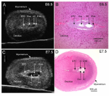
          In 40 MHz ultrasound images at E6.5, the ectoplacental cone region and proamniotic cavity could be visualized. The ectoplacental cone region was successfully targeted with 13.8 nL of a fluorescent bead suspension with few or no beads off-target in 51% of concepti microinjected at E6.5 (28/55 injected). Seventy eight percent of the embryos survived 2 to 12 days post injection (93/119), 73% (41/56) survived to term of which 68% (38/56) survived and appeared normal one week after birth. At E7.5, the amniotic and exocoelomic cavities, and ectoplacental cone region were discernable. Our success at targeting with few or no beads off-target was 90% (36/40) for the ectoplacental cone region and 81% (35/43) for the exocoelomic cavity but tended to be less, 68% (34/50), for the smaller amniotic cavity. At E11.5, beads microinjected at E7.5 into the ectoplacental cone region were found in the placental spongiotrophoblast layer, those injected into the exocoelomic cavity were found on the surface or within the placental labyrinth, and those injected into the amniotic cavity were found on the surface or within the embryo. Following microinjection at E7.5, survival one week after birth was 60% (26/43) when the amniotic cavity was the target and 66% (19/29) when the target was the ectoplacental cone region. The survival rate was similar in sham experiments, 54% (33/61), for which procedures were identical but no microinjection was performed, suggesting that surgery and manipulation of the uterus were the main causes of embryonic death.

          Conclusion

          Ultrasound-guided microinjection into the ectoplacental cone region at E6.5 or E7.5 and the amniotic cavity at E7.5 was achieved with a 7 day postnatal survival of ≥60%. Target accuracy of these sites and of the exocoelomic cavity at E7.5 was ≥51%. We suggest that this approach may be useful for exploring gene function during early placental and embryonic development.

          Related collections

          Most cited references26

          • Record: found
          • Abstract: found
          • Article: not found

          Development of structures and transport functions in the mouse placenta.

          The placenta is essential for sustaining the growth of the fetus during gestation, and defects in its function result in fetal growth restriction or, if more severe, fetal death. Several molecular pathways have been identified that are essential for development of the placenta, and mouse mutants offer new insights into the cell biology of placental development and physiology of nutrient transport.
            Bookmark
            • Record: found
            • Abstract: found
            • Article: not found

            What cardiovascular defect does my prenatal mouse mutant have, and why?

            Since the advent of mouse targeted mutations, gene traps, an escalating use of a variety of complex transgenic manipulations, and large-scale chemical mutagenesis projects yielding many mutants with cardiovascular defects, it has become increasingly evident that defects within the heart and vascular system are largely responsible for the observed in utero lethality of the embryo and early fetus. If a transgenically altered embryo survives implantation but fails to be born, it usually indicates that there is some form of lethal cardiovascular defect present. A number of embryonic organ and body systems, including the central nervous system, gut, lungs, urogenital system, and musculoskeletal system appear to have little or no survival value in utero (Copp, 1995). Cardiovascular abnormalities include the failure to establish an adequate yolk-sac vascular circulation, which results in early lethality (E8.5-10.5); poor cardiac function (E9.0-birth); failure to undergo correct looping and chamber formation of the primitive heart tube (E9.0-11.0); improper septation, including division of the common ventricle and atria and the establishment of a divided outflow tract (E11.0-13.0); inadequate establishment of the cardiac conduction system (E12.0-birth); and the failure of the in utero cardiovascular system to adapt to adult life (birth) and close the interatrial and aorta-pulmonary trunk shunts that are required for normal fetal life. Importantly, the developmental timing of lethality is usually a good indicator of both the type of the cardiovascular defect present and may also suggest the possible underlying cause/s. The purpose of this review is both to review the literature and to provide a beginner's guide for analysing cardiovascular defects in mouse mutants. Copyright 2002 Wiley-Liss, Inc.
              Bookmark
              • Record: found
              • Abstract: found
              • Article: not found

              40 MHz Doppler characterization of umbilical and dorsal aortic blood flow in the early mouse embryo.

              Physiological study of the developing mouse circulation has lagged behind advances in molecular cardiology. Using an innovative high-frequency Doppler system, we noninvasively characterized circulatory hemodynamics in early mouse embryos. We used image-guided 43 MHz pulsed-wave (PW) Doppler ultrasound to study the umbilical artery and vein, or dorsal aorta in 109 embryos. Studies were conducted on embryonic days (E) 9.5-14.5. Heart rate, peak blood flow velocities, and velocity time integrals in all vessels increased from E9.5-14.5, indicating increasing stroke volume and cardiac output. Heart rate, ranging from 192 bpm (E9.5) to 261 bpm (E14.5), was higher than previously reported. Placental impedance, assessed by the time delay between the peaks of the umbilical arterial and venous waveforms and by venous pulsatility, decreased with gestation. Acceleration time, a load-independent Doppler index of cardiac contractility, remained constant but seemed sensitive to heart rate. High-frequency PW Doppler is a powerful tool for the quantitative, noninvasive investigation of early mouse circulatory development.
                Bookmark

                Author and article information

                Journal
                BMC Dev Biol
                BMC Developmental Biology
                BioMed Central (London )
                1471-213X
                2006
                27 February 2006
                : 6
                : 10
                Affiliations
                [1 ]Samuel Lunenfeld Research Institute of Mount Sinai Hospital, Toronto, Canada
                [2 ]Department of Obstetrics and Gynecology, University of Toronto, Toronto, Canada
                [3 ]Department of Physiology, University of Toronto, Toronto, Canada
                [4 ]Department of Medical Genetics and Microbiology, University of Toronto, Toronto, Canada
                Article
                1471-213X-6-10
                10.1186/1471-213X-6-10
                1436015
                16504164
                d5c57864-a07b-44c6-975b-f3fa907b882c
                Copyright © 2006 Slevin et al; licensee BioMed Central Ltd.

                This is an Open Access article distributed under the terms of the Creative Commons Attribution License ( http://creativecommons.org/licenses/by/2.0), which permits unrestricted use, distribution, and reproduction in any medium, provided the original work is properly cited.

                History
                : 8 August 2005
                : 27 February 2006
                Categories
                Methodology Article

                Developmental biology
                Developmental biology

                Comments

                Comment on this article