3
views
0
recommends
+1 Recommend
0 collections
    0
    shares
      • Record: found
      • Abstract: found
      • Article: found
      Is Open Access

      TRIM22 activates NF-κB signaling in glioblastoma by accelerating the degradation of IκBα

      research-article

      Read this article at

      Bookmark
          There is no author summary for this article yet. Authors can add summaries to their articles on ScienceOpen to make them more accessible to a non-specialist audience.

          Abstract

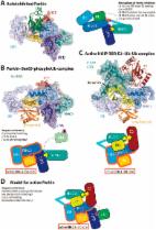
          NF-κB signaling plays a critical role in tumor growth and treatment resistance in GBM as in many other cancers. However, the molecular mechanisms underlying high, constitutive NF-κB activity in GBM remains to be elucidated. Here, we screened a panel of tripartite motif (TRIM) family proteins and identified TRIM22 as a potential activator of NF-κB using an NF-κB driven luciferase reporter construct in GBM cell lines. Knockout of TRIM22 using Cas9-sgRNAs led to reduced GBM cell proliferation, while TRIM22 overexpression enhanced proliferation of cell populations, in vitro and in an orthotopic xenograft model. However, two TRIM22 mutants, one with a critical RING-finger domain deletion and the other with amino acid changes at two active sites of RING E3 ligase (C15/18A), were both unable to promote GBM cell proliferation over controls, thus implicating E3 ligase activity in the growth-promoting properties of TRIM22. Co-immunoprecipitations demonstrated that TRIM22 bound a negative regulator of NF-κB, NF-κB inhibitor alpha (IκBα), and accelerated its degradation by inducing K48-linked ubiquitination. TRIM22 also formed a complex with the NF-κB upstream regulator IKKγ and promoted K63-linked ubiquitination, which led to the phosphorylation of both IKKα/β and IκBα. Expression of a non-phosphorylation mutant, srIκBα, inhibited the growth-promoting properties of TRIM22 in GBM cell lines. Finally, TRIM22 was increased in a cohort of primary GBM samples on a tissue microarray, and high expression of TRIM22 correlated with other clinical parameters associated with progressive gliomas, such as wild-type IDH1 status. In summary, our study revealed that TRIM22 activated NF-κB signaling through posttranslational modification of two critical regulators of NF-κB signaling in GBM cells.

          Related collections

          Most cited references51

          • Record: found
          • Abstract: found
          • Article: found

          Integrated Genomic Analysis Identifies Clinically Relevant Subtypes of Glioblastoma Characterized by Abnormalities in PDGFRA, IDH1, EGFR, and NF1

          The Cancer Genome Atlas Network recently cataloged recurrent genomic abnormalities in glioblastoma multiforme (GBM). We describe a robust gene expression-based molecular classification of GBM into Proneural, Neural, Classical, and Mesenchymal subtypes and integrate multidimensional genomic data to establish patterns of somatic mutations and DNA copy number. Aberrations and gene expression of EGFR, NF1, and PDGFRA/IDH1 each define the Classical, Mesenchymal, and Proneural subtypes, respectively. Gene signatures of normal brain cell types show a strong relationship between subtypes and different neural lineages. Additionally, response to aggressive therapy differs by subtype, with the greatest benefit in the Classical subtype and no benefit in the Proneural subtype. We provide a framework that unifies transcriptomic and genomic dimensions for GBM molecular stratification with important implications for future studies. Copyright (c) 2010 Elsevier Inc. All rights reserved.
            Bookmark
            • Record: found
            • Abstract: found
            • Article: not found

            Shared principles in NF-kappaB signaling.

            The transcription factor NF-kappaB has served as a standard for inducible transcription factors for more than 20 years. The numerous stimuli that activate NF-kappaB, and the large number of genes regulated by NF-kappaB, ensure that this transcription factor is still the subject of intense research. Here, we attempt to synthesize some of the basic principles that have emerged from studies of NF-kappaB, and we aim to generate a more unified view of NF-kappaB regulation.
              Bookmark
              • Record: found
              • Abstract: found
              • Article: found
              Is Open Access

              Ubiquitin modifications

              Protein ubiquitination is a dynamic multifaceted post-translational modification involved in nearly all aspects of eukaryotic biology. Once attached to a substrate, the 76-amino acid protein ubiquitin is subjected to further modifications, creating a multitude of distinct signals with distinct cellular outcomes, referred to as the 'ubiquitin code'. Ubiquitin can be ubiquitinated on seven lysine (Lys) residues or on the N-terminus, leading to polyubiquitin chains that can encompass complex topologies. Alternatively or in addition, ubiquitin Lys residues can be modified by ubiquitin-like molecules (such as SUMO or NEDD8). Finally, ubiquitin can also be acetylated on Lys, or phosphorylated on Ser, Thr or Tyr residues, and each modification has the potential to dramatically alter the signaling outcome. While the number of distinctly modified ubiquitin species in cells is mind-boggling, much progress has been made to characterize the roles of distinct ubiquitin modifications, and many enzymes and receptors have been identified that create, recognize or remove these ubiquitin modifications. We here provide an overview of the various ubiquitin modifications present in cells, and highlight recent progress on ubiquitin chain biology. We then discuss the recent findings in the field of ubiquitin acetylation and phosphorylation, with a focus on Ser65-phosphorylation and its role in mitophagy and Parkin activation.
                Bookmark

                Author and article information

                Contributors
                hb@sdu.edu.cn
                lixg@sdu.edu.cn
                jian.wang@uib.no
                Journal
                Cell Death Differ
                Cell Death Differ
                Cell Death and Differentiation
                Nature Publishing Group UK (London )
                1350-9047
                1476-5403
                19 August 2020
                19 August 2020
                January 2021
                : 28
                : 1
                : 367-381
                Affiliations
                [1 ]GRID grid.27255.37, ISNI 0000 0004 1761 1174, Department of Neurosurgery, Qilu Hospital and Institute of Brain and Brain-Inspired Science, Cheeloo College of Medicine, , Shandong University, ; Jinan, China
                [2 ]Shandong Key Laboratory of Brain Function Remodeling, Jinan, China
                [3 ]GRID grid.7914.b, ISNI 0000 0004 1936 7443, Department of Biomedicine, , University of Bergen, ; Jonas Lies vei 91, 5009 Bergen, Norway
                [4 ]GRID grid.7914.b, ISNI 0000 0004 1936 7443, Molecular Imaging Center, , University of Bergen, ; Jonas Lies vei 91, 5009 Bergen, Norway
                Author information
                http://orcid.org/0000-0002-9482-5227
                Article
                606
                10.1038/s41418-020-00606-w
                7853150
                32814880
                d624acbe-83e2-4544-970f-19b1c228b499
                © The Author(s) 2020

                Open Access This article is licensed under a Creative Commons Attribution 4.0 International License, which permits use, sharing, adaptation, distribution and reproduction in any medium or format, as long as you give appropriate credit to the original author(s) and the source, provide a link to the Creative Commons license, and indicate if changes were made. The images or other third party material in this article are included in the article’s Creative Commons license, unless indicated otherwise in a credit line to the material. If material is not included in the article’s Creative Commons license and your intended use is not permitted by statutory regulation or exceeds the permitted use, you will need to obtain permission directly from the copyright holder. To view a copy of this license, visit http://creativecommons.org/licenses/by/4.0/.

                History
                : 25 December 2019
                : 29 July 2020
                : 6 August 2020
                Funding
                Funded by: FundRef https://doi.org/10.13039/501100001809, National Natural Science Foundation of China (National Science Foundation of China);
                Award ID: 81972351
                Award ID: 81903126
                Award ID: 81701329
                Award ID: 81874083
                Award ID: 81702474
                Award Recipient :
                Funded by: FundRef https://doi.org/10.13039/100012905, Department of Science and Technology of Shandong Province;
                Award ID: 2017CXGC1502
                Award ID: 2018CXGC1503
                Award ID: 2017CXGC1203
                Award ID: 2018GSF118082
                Award Recipient :
                Funded by: the Special Foundation for Taishan Scholars tshw201502056 the Shandong Provincial Natural Science Foundation ZR2017MH116 the Jinan Science and Technology Bureau of Shandong Province 2019GXRC006
                Funded by: FundRef https://doi.org/10.13039/501100002858, China Postdoctoral Science Foundation;
                Award ID: 2018M642666
                Award Recipient :
                Funded by: the Special Foundation for Taishan Scholars tsqn201909173
                Funded by: the Special Foundation for Taishan Scholars tsqn20161067 the Shandong Provincial Natural Science Foundation ZR2017MH015
                Funded by: the Special Foundation for Taishan Scholars ts20110814
                Categories
                Article
                Custom metadata
                © ADMC Associazione Differenziamento e Morte Cellulare 2021

                Cell biology
                cns cancer,oncogenes,ubiquitin ligases
                Cell biology
                cns cancer, oncogenes, ubiquitin ligases

                Comments

                Comment on this article